Magnesium »
PDB 8hhc-8htg »
8hls »
Magnesium in PDB 8hls: Crystal Structure of the Sialidase AM1547 From A.Muciniphila
Protein crystallography data
The structure of Crystal Structure of the Sialidase AM1547 From A.Muciniphila, PDB code: 8hls
was solved by
B.Rui,
T.Xinyue,
with X-Ray Crystallography technique. A brief refinement statistics is given in the table below:
| Resolution Low / High (Å) | 21.64 / 2.04 |
| Space group | P 1 21 1 |
| Cell size a, b, c (Å), α, β, γ (°) | 72.16, 56.771, 145.932, 90, 94.01, 90 |
| R / Rfree (%) | 18 / 22.3 |
Other elements in 8hls:
The structure of Crystal Structure of the Sialidase AM1547 From A.Muciniphila also contains other interesting chemical elements:
| Chlorine | (Cl) | 4 atoms |
Magnesium Binding Sites:
The binding sites of Magnesium atom in the Crystal Structure of the Sialidase AM1547 From A.Muciniphila
(pdb code 8hls). This binding sites where shown within
5.0 Angstroms radius around Magnesium atom.
In total 2 binding sites of Magnesium where determined in the Crystal Structure of the Sialidase AM1547 From A.Muciniphila, PDB code: 8hls:
Jump to Magnesium binding site number: 1; 2;
In total 2 binding sites of Magnesium where determined in the Crystal Structure of the Sialidase AM1547 From A.Muciniphila, PDB code: 8hls:
Jump to Magnesium binding site number: 1; 2;
Magnesium binding site 1 out of 2 in 8hls
Go back to
Magnesium binding site 1 out
of 2 in the Crystal Structure of the Sialidase AM1547 From A.Muciniphila
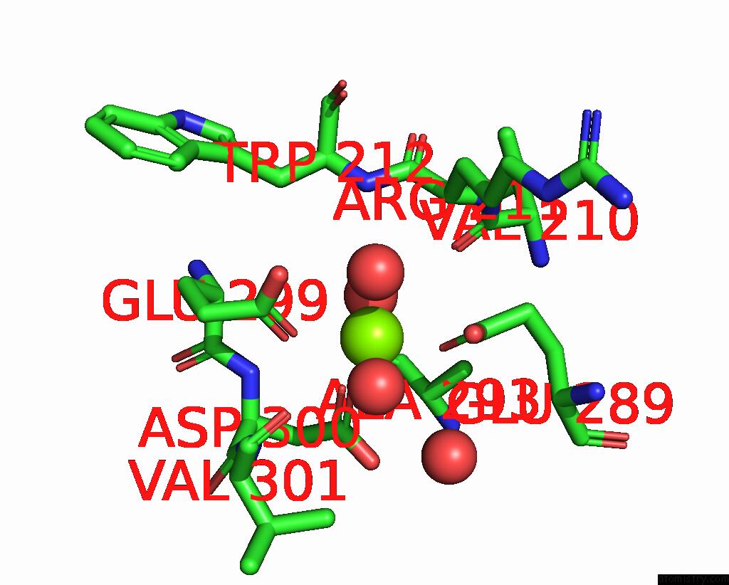
Mono view
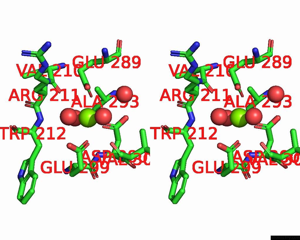
Stereo pair view
Mono view
Stereo pair view
A full contact list of Magnesium with other atoms in the Mg binding
site number 1 of Crystal Structure of the Sialidase AM1547 From A.Muciniphila within 5.0Å range:
|
Magnesium binding site 2 out of 2 in 8hls
Go back to
Magnesium binding site 2 out
of 2 in the Crystal Structure of the Sialidase AM1547 From A.Muciniphila
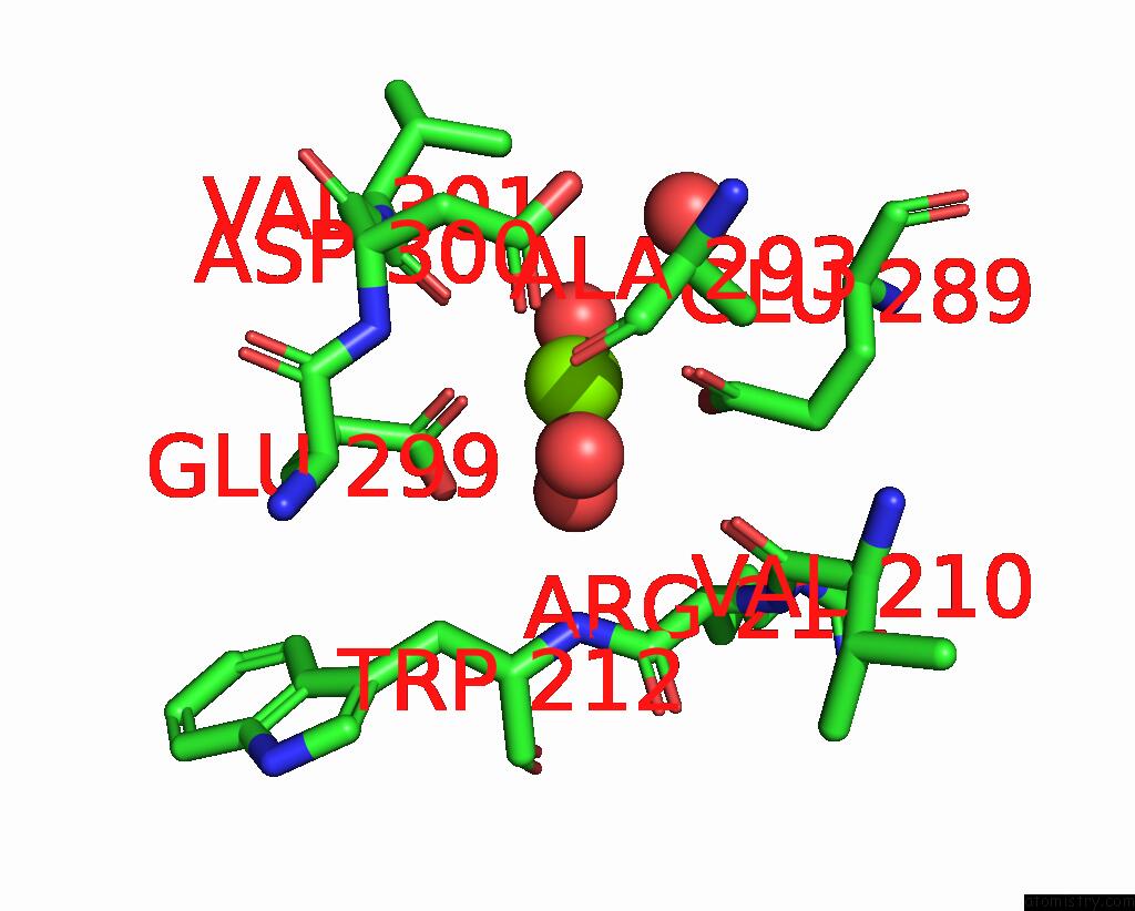
Mono view
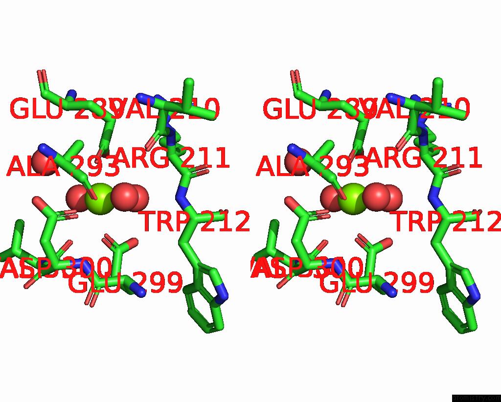
Stereo pair view
Mono view
Stereo pair view
A full contact list of Magnesium with other atoms in the Mg binding
site number 2 of Crystal Structure of the Sialidase AM1547 From A.Muciniphila within 5.0Å range:
|
Reference:
T.Xinyue,
B.Rui.
Structural Analysis of A Novel Sialidase From Akkermansia Muciniphila Reveals A Distinct Catalytic Motif in GH33 Family To Be Published.
Page generated: Fri Aug 15 06:35:51 2025
Last articles
Mn in 9LJUMn in 9LJW
Mn in 9LJS
Mn in 9LJR
Mn in 9LJT
Mn in 9LJV
Mg in 9UA2
Mg in 9R96
Mg in 9VM1
Mg in 9P01