Atomistry » Magnesium » PDB 8hhb-8hsr » 8hmu
Atomistry »
  Magnesium »
    PDB 8hhb-8hsr »
      8hmu »

Magnesium in PDB 8hmu: Crystal Structure of PKM2 Mutant R516C

Enzymatic activity of Crystal Structure of PKM2 Mutant R516C

All present enzymatic activity of Crystal Structure of PKM2 Mutant R516C:
2.7.1.40; 2.7.10.2; 2.7.11.1;

Protein crystallography data

The structure of Crystal Structure of PKM2 Mutant R516C, PDB code: 8hmu was solved by S.Upadhyay, A.Kumar, A.K.Patel, with X-Ray Crystallography technique. A brief refinement statistics is given in the table below:

Resolution Low / High (Å) 49.92 / 2.50
Space group P 21 21 21
Cell size a, b, c (Å), α, β, γ (°) 105.4, 138.512, 155.614, 90, 90, 90
R / Rfree (%) 23.7 / 27.9

Magnesium Binding Sites:

The binding sites of Magnesium atom in the Crystal Structure of PKM2 Mutant R516C (pdb code 8hmu). This binding sites where shown within 5.0 Angstroms radius around Magnesium atom.
In total 7 binding sites of Magnesium where determined in the Crystal Structure of PKM2 Mutant R516C, PDB code: 8hmu:
Jump to Magnesium binding site number: 1; 2; 3; 4; 5; 6; 7;

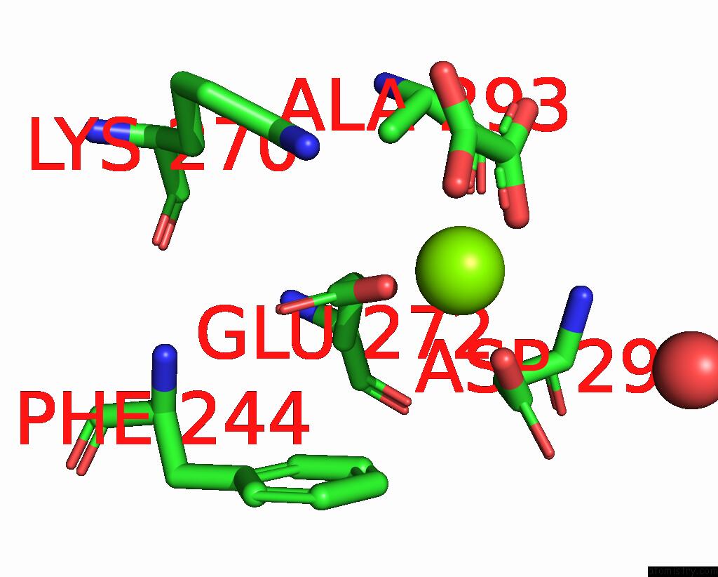
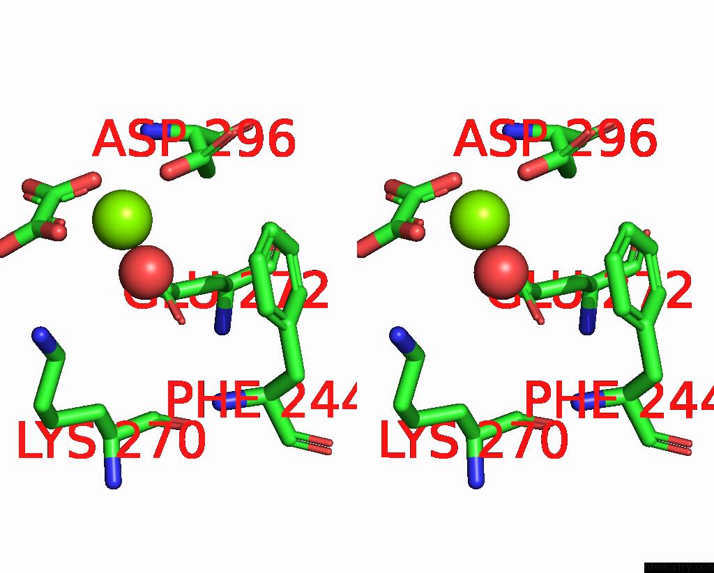
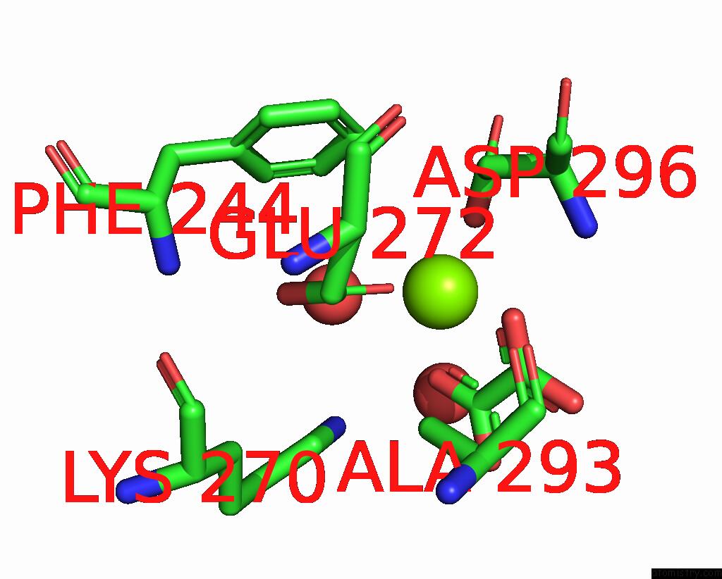
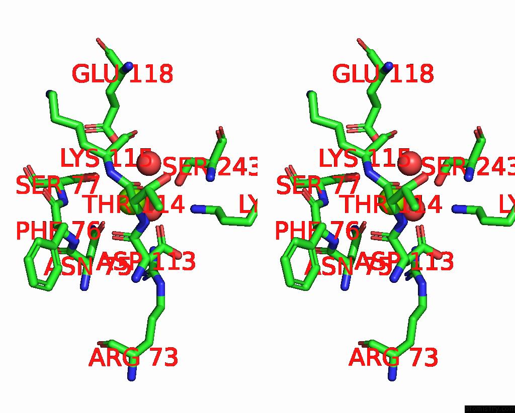
Magnesium binding site 1 out of 7 in 8hmu

Go back to Magnesium Binding Sites List in 8hmu
Magnesium binding site 1 out of 7 in the Crystal Structure of PKM2 Mutant R516C


Mono view


Stereo pair view

A full contact list of Magnesium with other atoms in the Mg binding site number 1 of Crystal Structure of PKM2 Mutant R516C within 5.0Å range:
probe atom residue distance (Å) B Occ
A:Mg607

b:40.9
occ:1.00
OE2 A:GLU272 1.9 44.9 1.0
O1 A:OXL602 1.9 47.8 1.0
O2 A:OXL602 2.1 44.6 1.0
OD2 A:ASP296 2.1 45.2 1.0
C1 A:OXL602 2.7 48.3 1.0
C2 A:OXL602 2.8 49.1 1.0
CD A:GLU272 3.1 44.3 1.0
CG A:ASP296 3.3 47.5 1.0
O3 A:OXL602 3.8 49.2 1.0
NZ A:LYS270 3.9 43.5 1.0
OE1 A:GLU272 4.0 42.8 1.0
CB A:ASP296 4.0 43.3 1.0
O4 A:OXL602 4.0 47.0 1.0
CG A:GLU272 4.0 43.9 1.0
OD1 A:ASP296 4.3 48.8 1.0
N A:ASP296 4.4 45.8 1.0
CZ A:PHE244 4.5 51.7 1.0
CE A:LYS270 4.5 45.3 1.0
CE1 A:PHE244 4.8 48.1 1.0
CB A:ALA293 4.8 40.5 1.0
CA A:ASP296 4.8 45.2 1.0
O A:HOH759 5.0 50.7 1.0

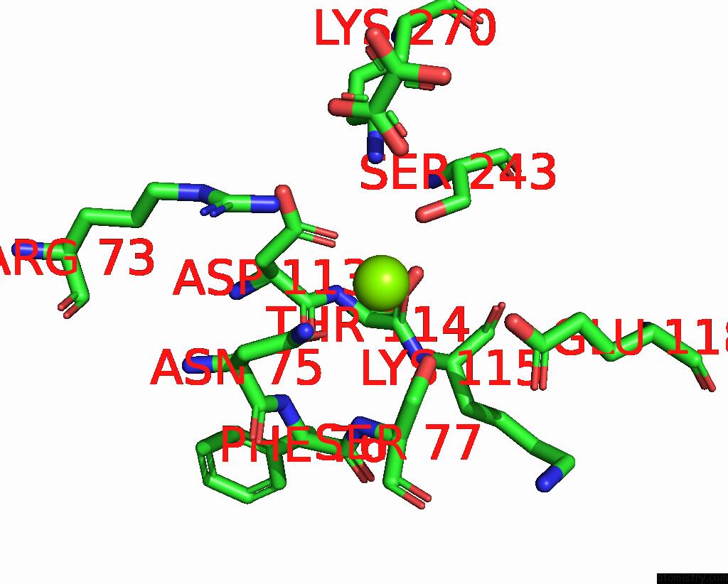
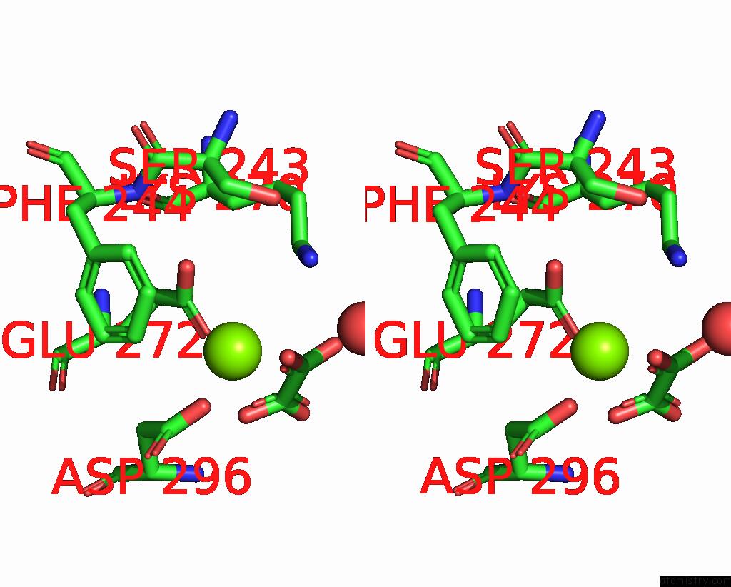
Magnesium binding site 2 out of 7 in 8hmu

Go back to Magnesium Binding Sites List in 8hmu
Magnesium binding site 2 out of 7 in the Crystal Structure of PKM2 Mutant R516C


Mono view


Stereo pair view

A full contact list of Magnesium with other atoms in the Mg binding site number 2 of Crystal Structure of PKM2 Mutant R516C within 5.0Å range:
probe atom residue distance (Å) B Occ
A:Mg608

b:41.1
occ:1.00
OD1 A:ASP113 2.5 42.4 1.0
OD1 A:ASN75 2.7 43.0 1.0
O A:THR114 2.9 49.7 1.0
OG A:SER77 2.9 46.6 1.0
CG A:ASP113 3.5 43.2 1.0
OG A:SER243 3.6 42.0 1.0
CG A:ASN75 3.8 44.8 1.0
C A:THR114 3.8 49.0 1.0
CB A:SER77 4.0 51.0 1.0
NZ A:LYS270 4.0 43.5 1.0
O A:ASP113 4.1 42.5 1.0
CA A:LYS115 4.2 49.1 1.0
O A:LYS115 4.2 49.5 1.0
ND2 A:ASN75 4.3 54.7 1.0
CB A:ASP113 4.3 43.6 1.0
OD2 A:ASP113 4.3 42.7 1.0
NH2 A:ARG73 4.3 40.5 1.0
C A:ASP113 4.3 43.9 1.0
N A:LYS115 4.4 46.2 1.0
N A:SER77 4.4 48.2 1.0
C A:LYS115 4.6 47.8 1.0
O1 A:OXL602 4.7 47.8 1.0
CB A:SER243 4.7 44.6 1.0
OE2 A:GLU118 4.7 53.2 1.0
N A:THR114 4.7 48.2 1.0
CA A:SER77 4.8 48.2 1.0
CA A:THR114 4.9 47.8 1.0
CA A:ASP113 4.9 44.5 1.0
CB A:ASN75 5.0 39.5 1.0
N A:PHE76 5.0 46.0 1.0

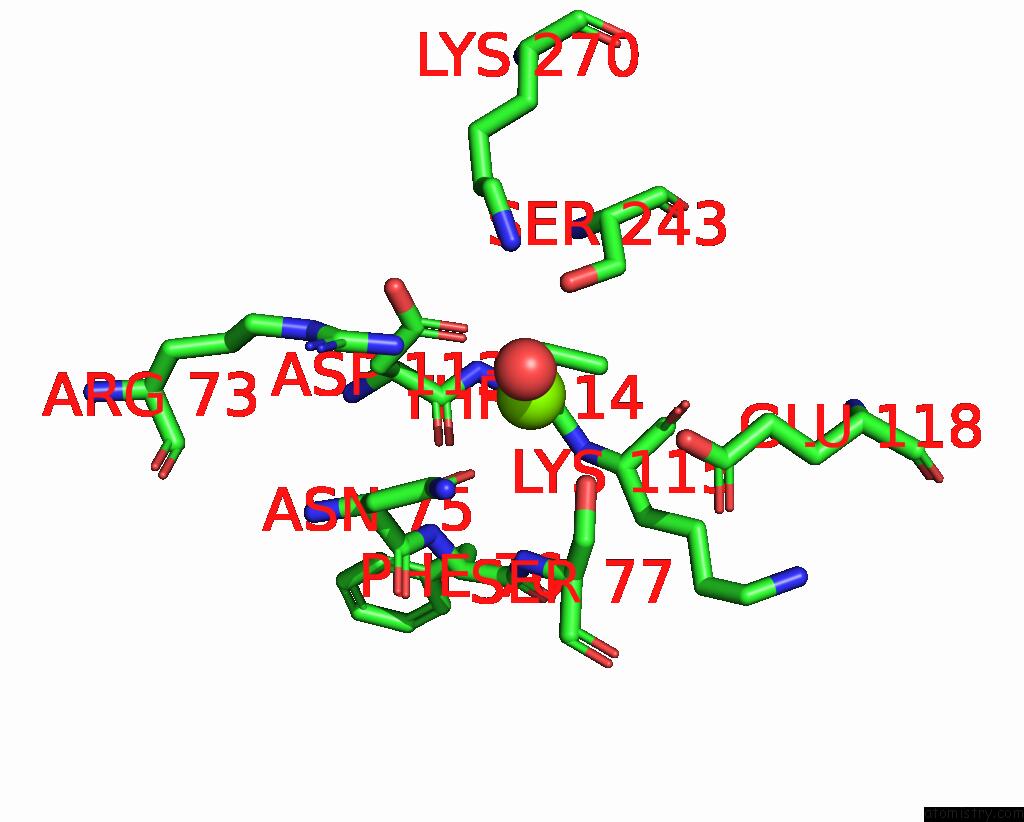
Magnesium binding site 3 out of 7 in 8hmu

Go back to Magnesium Binding Sites List in 8hmu
Magnesium binding site 3 out of 7 in the Crystal Structure of PKM2 Mutant R516C


Mono view


Stereo pair view

A full contact list of Magnesium with other atoms in the Mg binding site number 3 of Crystal Structure of PKM2 Mutant R516C within 5.0Å range:
probe atom residue distance (Å) B Occ
B:Mg609

b:63.5
occ:1.00
OE2 B:GLU272 1.9 55.4 1.0
O3 B:OXL602 1.9 62.4 1.0
OD2 B:ASP296 2.0 61.7 1.0
O B:HOH746 2.4 62.7 1.0
O4 B:OXL602 2.7 57.2 1.0
C1 B:OXL602 2.9 59.6 1.0
CD B:GLU272 3.0 62.5 1.0
CG B:ASP296 3.2 61.8 1.0
C2 B:OXL602 3.3 57.1 1.0
OE1 B:GLU272 3.5 61.7 1.0
CB B:ASP296 3.9 58.1 1.0
O1 B:OXL602 4.1 57.2 1.0
NZ B:LYS270 4.1 55.0 1.0
OD1 B:ASP296 4.1 65.3 1.0
CG B:GLU272 4.2 61.8 1.0
CE1 B:PHE244 4.3 66.5 1.0
CZ B:PHE244 4.3 68.9 1.0
O2 B:OXL602 4.5 53.5 1.0
CE B:LYS270 4.6 57.0 1.0
N B:ASP296 4.6 55.8 1.0
CA B:ASP296 4.9 59.4 1.0

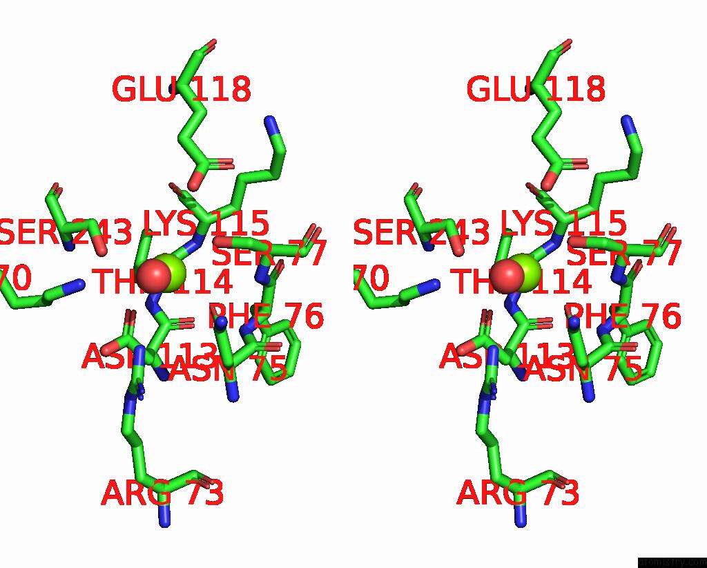
Magnesium binding site 4 out of 7 in 8hmu

Go back to Magnesium Binding Sites List in 8hmu
Magnesium binding site 4 out of 7 in the Crystal Structure of PKM2 Mutant R516C


Mono view


Stereo pair view

A full contact list of Magnesium with other atoms in the Mg binding site number 4 of Crystal Structure of PKM2 Mutant R516C within 5.0Å range:
probe atom residue distance (Å) B Occ
C:Mg608

b:46.5
occ:1.00
OE1 C:GLU272 1.9 51.1 1.0
OD2 C:ASP296 1.9 53.8 1.0
O1 C:OXL602 2.1 45.8 1.0
O2 C:OXL602 2.3 46.8 1.0
C1 C:OXL602 2.8 47.0 1.0
C2 C:OXL602 2.8 46.3 1.0
CD C:GLU272 3.1 51.9 1.0
CG C:ASP296 3.1 52.2 1.0
OE2 C:GLU272 3.7 51.0 1.0
CB C:ASP296 3.8 49.8 1.0
O C:HOH736 4.0 48.1 1.0
O3 C:OXL602 4.1 48.2 1.0
OD1 C:ASP296 4.1 55.8 1.0
NZ C:LYS270 4.1 44.8 1.0
CE2 C:PHE244 4.1 64.8 1.0
O4 C:OXL602 4.1 44.8 1.0
CZ C:PHE244 4.1 62.9 1.0
O C:HOH748 4.2 53.1 1.0
CG C:GLU272 4.3 50.7 1.0
N C:ASP296 4.4 44.7 1.0
CE C:LYS270 4.6 48.5 1.0
CB C:ALA293 4.7 43.4 1.0
CB C:GLU272 4.8 51.9 1.0
CA C:ASP296 4.8 48.4 1.0
CD2 C:PHE244 5.0 63.6 1.0

Magnesium binding site 5 out of 7 in 8hmu

Go back to Magnesium Binding Sites List in 8hmu
Magnesium binding site 5 out of 7 in the Crystal Structure of PKM2 Mutant R516C


Mono view


Stereo pair view

A full contact list of Magnesium with other atoms in the Mg binding site number 5 of Crystal Structure of PKM2 Mutant R516C within 5.0Å range:
probe atom residue distance (Å) B Occ
C:Mg609

b:42.8
occ:1.00
OD1 C:ASN75 2.5 51.1 1.0
OD1 C:ASP113 2.6 45.2 1.0
O C:HOH748 2.6 53.1 1.0
O C:THR114 2.7 48.3 1.0
OG C:SER77 2.9 53.8 1.0
O C:HOH736 3.1 48.1 1.0
CG C:ASP113 3.6 45.8 1.0
OG C:SER243 3.6 47.1 1.0
CG C:ASN75 3.6 50.3 1.0
C C:THR114 3.7 49.0 1.0
O C:ASP113 4.0 46.8 1.0
NZ C:LYS270 4.0 44.8 1.0
NH2 C:ARG73 4.0 44.3 1.0
CB C:SER77 4.1 52.8 1.0
CA C:LYS115 4.2 50.8 1.0
C C:ASP113 4.2 46.4 1.0
CB C:ASP113 4.2 44.2 1.0
N C:LYS115 4.3 50.0 1.0
N C:SER77 4.3 52.4 1.0
ND2 C:ASN75 4.3 53.0 1.0
OE2 C:GLU118 4.3 64.6 1.0
N C:THR114 4.5 49.7 1.0
O C:LYS115 4.5 56.0 1.0
OD2 C:ASP113 4.5 45.7 1.0
N C:PHE76 4.6 47.3 1.0
CA C:THR114 4.7 48.7 1.0
C C:LYS115 4.7 54.2 1.0
CA C:SER77 4.7 53.2 1.0
CB C:SER243 4.7 50.9 1.0
CB C:ASN75 4.8 45.2 1.0
CA C:ASN75 4.8 44.8 1.0
CA C:ASP113 4.8 46.4 1.0
C C:ASN75 5.0 47.9 1.0

Magnesium binding site 6 out of 7 in 8hmu

Go back to Magnesium Binding Sites List in 8hmu
Magnesium binding site 6 out of 7 in the Crystal Structure of PKM2 Mutant R516C


Mono view


Stereo pair view

A full contact list of Magnesium with other atoms in the Mg binding site number 6 of Crystal Structure of PKM2 Mutant R516C within 5.0Å range:
probe atom residue distance (Å) B Occ
D:Mg609

b:40.4
occ:1.00
O3 D:OXL602 1.8 37.2 1.0
OE1 D:GLU272 1.9 47.3 1.0
OD2 D:ASP296 1.9 38.9 1.0
CD D:GLU272 2.8 47.2 1.0
C1 D:OXL602 2.8 39.8 1.0
O2 D:OXL602 3.0 38.0 1.0
OE2 D:GLU272 3.0 46.8 1.0
CG D:ASP296 3.1 39.4 1.0
CZ D:PHE244 3.4 46.1 1.0
C2 D:OXL602 3.5 40.3 1.0
NZ D:LYS270 3.8 39.6 1.0
CB D:ASP296 3.8 39.6 1.0
CE2 D:PHE244 3.9 46.8 1.0
O1 D:OXL602 3.9 36.3 1.0
CE1 D:PHE244 4.0 40.2 1.0
OD1 D:ASP296 4.0 34.5 1.0
CG D:GLU272 4.2 40.5 1.0
O D:HOH775 4.3 37.0 1.0
CB D:GLU272 4.6 42.3 1.0
CE D:LYS270 4.6 40.2 1.0
O4 D:OXL602 4.7 38.3 1.0
CD2 D:PHE244 4.7 44.6 1.0
N D:ASP296 4.7 37.4 1.0
OG D:SER243 4.8 41.3 1.0
CD1 D:PHE244 4.8 41.6 1.0
CA D:ASP296 4.9 41.5 1.0

Magnesium binding site 7 out of 7 in 8hmu

Go back to Magnesium Binding Sites List in 8hmu
Magnesium binding site 7 out of 7 in the Crystal Structure of PKM2 Mutant R516C


Mono view


Stereo pair view

A full contact list of Magnesium with other atoms in the Mg binding site number 7 of Crystal Structure of PKM2 Mutant R516C within 5.0Å range:
probe atom residue distance (Å) B Occ
D:Mg610

b:37.9
occ:1.00
O D:HOH775 2.4 37.0 1.0
O D:THR114 2.4 42.9 1.0
OD1 D:ASP113 2.6 42.8 1.0
OD1 D:ASN75 2.7 40.4 1.0
OG D:SER77 2.7 48.5 1.0
C D:THR114 3.5 42.2 1.0
OG D:SER243 3.5 41.3 1.0
CG D:ASP113 3.6 41.6 1.0
CG D:ASN75 3.8 40.7 1.0
NZ D:LYS270 3.9 39.6 1.0
CB D:SER77 4.0 48.4 1.0
NH2 D:ARG73 4.1 36.4 1.0
O D:ASP113 4.1 41.2 1.0
CA D:LYS115 4.2 46.4 1.0
C D:ASP113 4.2 41.5 1.0
N D:LYS115 4.3 47.7 1.0
O D:LYS115 4.3 47.8 1.0
N D:THR114 4.3 45.9 1.0
N D:SER77 4.3 45.4 1.0
CB D:ASP113 4.3 40.0 1.0
ND2 D:ASN75 4.4 41.2 1.0
OD2 D:ASP113 4.4 40.8 1.0
OE2 D:GLU118 4.5 56.1 1.0
CA D:THR114 4.6 42.9 1.0
C D:LYS115 4.6 46.6 1.0
CB D:SER243 4.6 40.9 1.0
CA D:SER77 4.7 46.1 1.0
N D:PHE76 4.8 46.2 1.0
CA D:ASP113 4.9 38.6 1.0
CB D:ASN75 4.9 40.1 1.0
CA D:ASN75 5.0 42.1 1.0

Reference:

S.Upadhyay, A.Kumar, A.K.Patel. Structural and Mechanistic Insights Into Cancer Patient-Derived Mutations in Pyruvate Kinase Muscle Isoform 2 To Be Published.
Page generated: Fri Aug 15 06:43:50 2025

Last articles

Mg in 8S8G
Mg in 8S8D
Mg in 8S8E
Mg in 8S8F
Mg in 8S1P
Mg in 8RW1
Mg in 8S87
Mg in 8S8B
Mg in 8S8A
Mg in 8S7G
© Copyright 2008-2020 by atomistry.com
Home   |    Site Map   |    Copyright   |    Contact us   |    Privacy