Magnesium »
PDB 8hh9-8hsj »
8hq9 »
Magnesium in PDB 8hq9: Crystal Structure of the Mlad Domain of the Mlad Protein From Escherichia Coli (Form II)
Protein crystallography data
The structure of Crystal Structure of the Mlad Domain of the Mlad Protein From Escherichia Coli (Form II), PDB code: 8hq9
was solved by
A.Dutta,
S.P.Kanaujia,
with X-Ray Crystallography technique. A brief refinement statistics is given in the table below:
| Resolution Low / High (Å) | 78.98 / 2.70 |
| Space group | P 21 2 21 |
| Cell size a, b, c (Å), α, β, γ (°) | 60.42, 106.95, 117.12, 90, 90, 90 |
| R / Rfree (%) | 20.4 / 26.1 |
Magnesium Binding Sites:
The binding sites of Magnesium atom in the Crystal Structure of the Mlad Domain of the Mlad Protein From Escherichia Coli (Form II)
(pdb code 8hq9). This binding sites where shown within
5.0 Angstroms radius around Magnesium atom.
In total only one binding site of Magnesium was determined in the Crystal Structure of the Mlad Domain of the Mlad Protein From Escherichia Coli (Form II), PDB code: 8hq9:
In total only one binding site of Magnesium was determined in the Crystal Structure of the Mlad Domain of the Mlad Protein From Escherichia Coli (Form II), PDB code: 8hq9:
Magnesium binding site 1 out of 1 in 8hq9
Go back to
Magnesium binding site 1 out
of 1 in the Crystal Structure of the Mlad Domain of the Mlad Protein From Escherichia Coli (Form II)
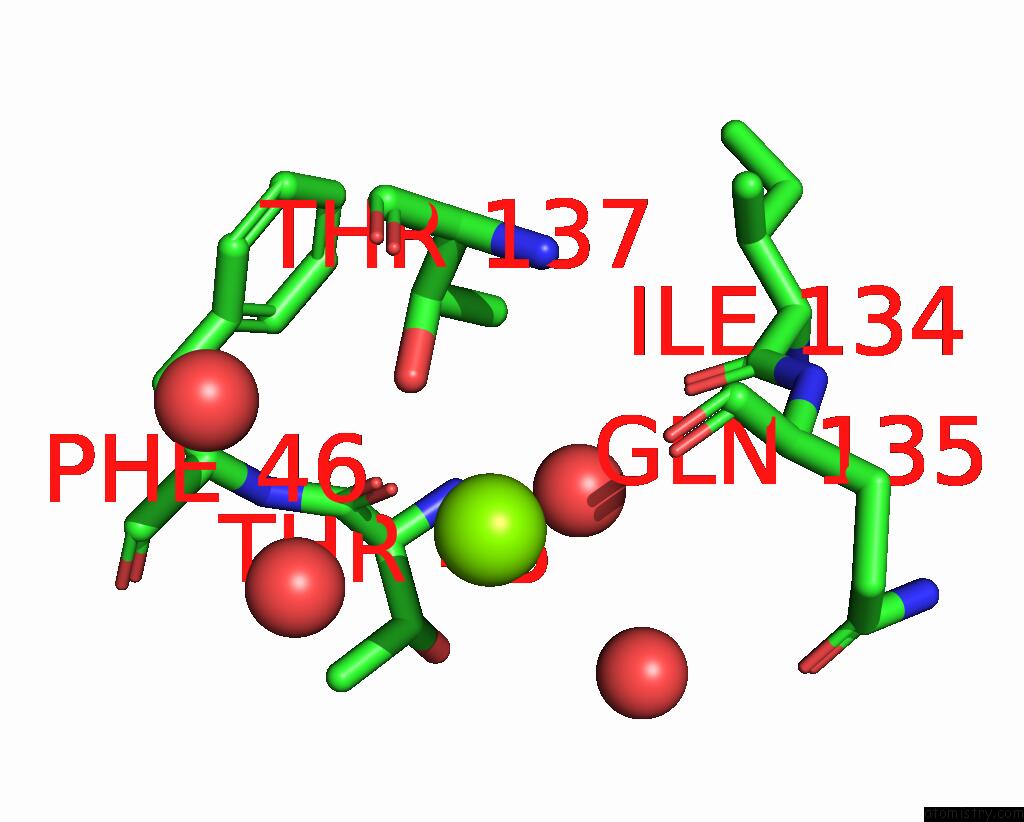
Mono view
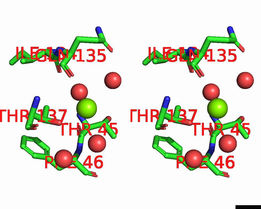
Stereo pair view
Mono view
Stereo pair view
A full contact list of Magnesium with other atoms in the Mg binding
site number 1 of Crystal Structure of the Mlad Domain of the Mlad Protein From Escherichia Coli (Form II) within 5.0Å range:
|
Reference:
A.Dutta,
S.P.Kanaujia.
The Structural Features of Mlad Illuminate Its Unique Ligand-Transporting Mechanism and Ancestry To Be Published.
Page generated: Fri Oct 4 08:12:57 2024
Last articles
Fe in 2YXOFe in 2YRS
Fe in 2YXC
Fe in 2YNM
Fe in 2YVJ
Fe in 2YP1
Fe in 2YU2
Fe in 2YU1
Fe in 2YQB
Fe in 2YOO