Magnesium »
PDB 8rav-8rjw »
8rav »
Magnesium in PDB 8rav: The Crystal Structure of Dna-Bound Human Mutsbeta (MSH2/MSH3) in the Canonical Mismatch Bound Conformation with Adp Bound in MSH2
Protein crystallography data
The structure of The Crystal Structure of Dna-Bound Human Mutsbeta (MSH2/MSH3) in the Canonical Mismatch Bound Conformation with Adp Bound in MSH2, PDB code: 8rav
was solved by
M.Thomsen,
E.Costanzi,
with X-Ray Crystallography technique. A brief refinement statistics is given in the table below:
| Resolution Low / High (Å) | 44.94 / 2.63 |
| Space group | P 1 |
| Cell size a, b, c (Å), α, β, γ (°) | 66.599, 91.12, 96.482, 67.56, 86.81, 74.64 |
| R / Rfree (%) | 23.9 / 28.4 |
Other elements in 8rav:
The structure of The Crystal Structure of Dna-Bound Human Mutsbeta (MSH2/MSH3) in the Canonical Mismatch Bound Conformation with Adp Bound in MSH2 also contains other interesting chemical elements:
| Sodium | (Na) | 1 atom |
Magnesium Binding Sites:
The binding sites of Magnesium atom in the The Crystal Structure of Dna-Bound Human Mutsbeta (MSH2/MSH3) in the Canonical Mismatch Bound Conformation with Adp Bound in MSH2
(pdb code 8rav). This binding sites where shown within
5.0 Angstroms radius around Magnesium atom.
In total only one binding site of Magnesium was determined in the The Crystal Structure of Dna-Bound Human Mutsbeta (MSH2/MSH3) in the Canonical Mismatch Bound Conformation with Adp Bound in MSH2, PDB code: 8rav:
In total only one binding site of Magnesium was determined in the The Crystal Structure of Dna-Bound Human Mutsbeta (MSH2/MSH3) in the Canonical Mismatch Bound Conformation with Adp Bound in MSH2, PDB code: 8rav:
Magnesium binding site 1 out of 1 in 8rav
Go back to
Magnesium binding site 1 out
of 1 in the The Crystal Structure of Dna-Bound Human Mutsbeta (MSH2/MSH3) in the Canonical Mismatch Bound Conformation with Adp Bound in MSH2
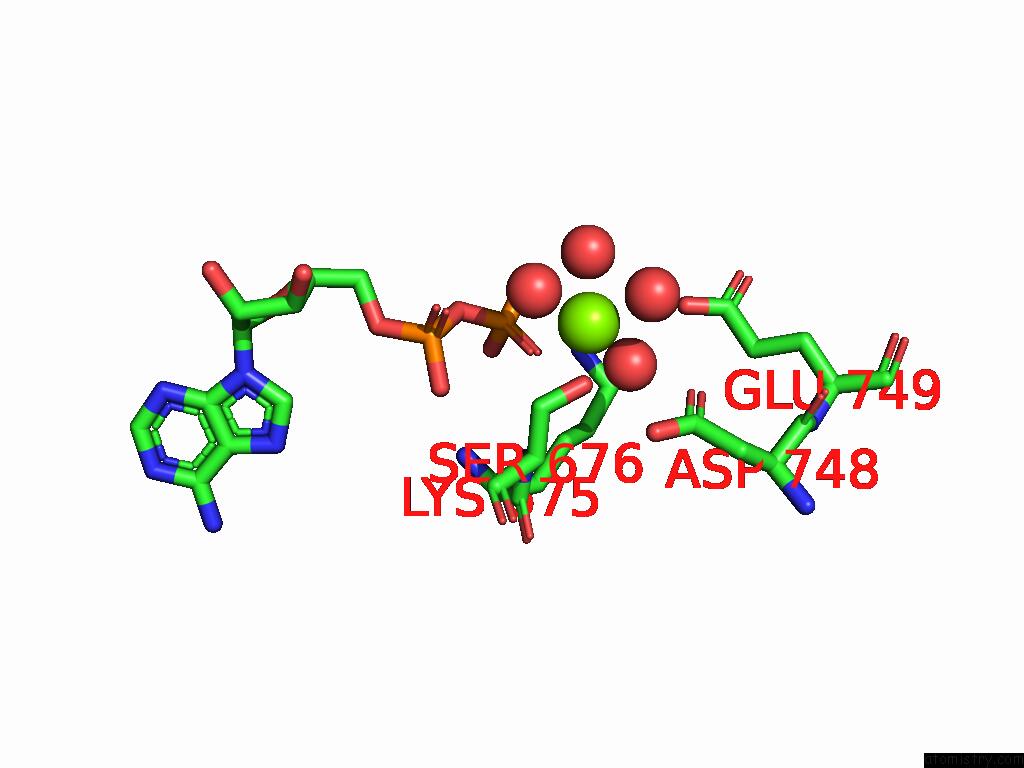
Mono view
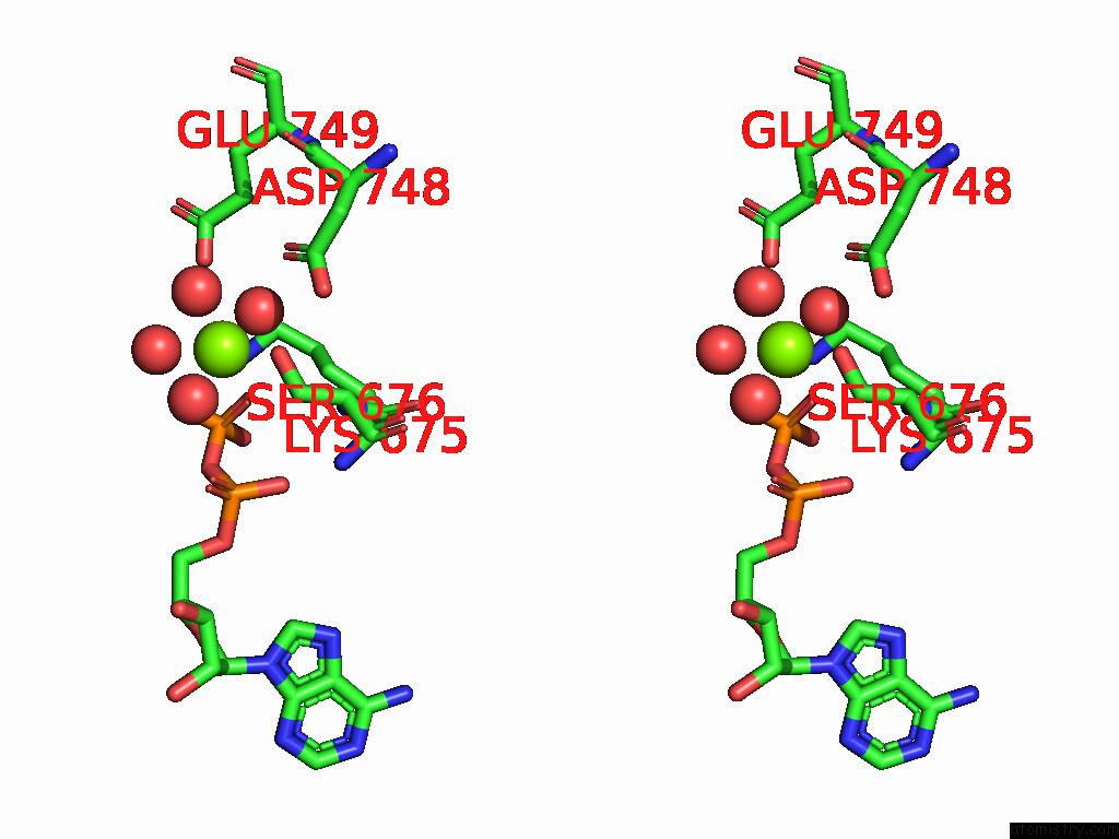
Stereo pair view
Mono view
Stereo pair view
A full contact list of Magnesium with other atoms in the Mg binding
site number 1 of The Crystal Structure of Dna-Bound Human Mutsbeta (MSH2/MSH3) in the Canonical Mismatch Bound Conformation with Adp Bound in MSH2 within 5.0Å range:
|
Reference:
M.Thomsen,
T.Neudegger,
G.Thieulin-Pardo,
M.Blaesse,
E.Costanzi,
S.Steinbacher,
N.V.Plotnikov,
C.Dominguez,
R.R.Iyer,
H.A.Wilkinson,
E.Monteagudo,
T.S.Haque,
B.C.Prasad,
M.Finley,
J.Boudet,
T.F.Vogt,
D.P.Felsenfeld.
The Crystal Structure of Dna-Bound Human Mutsbeta (MSH2/MSH3) in the Canonical Mismatch Bound Conformation with Adp Bound in MSH2 and MSH3 To Be Published.
Page generated: Fri Aug 15 13:56:01 2025
Last articles
Mg in 8V2JMg in 8UZ1
Mg in 8V1Q
Mg in 8V1S
Mg in 8UZ0
Mg in 8V0Y
Mg in 8V0K
Mg in 8V0I
Mg in 8V01
Mg in 8UZY