Magnesium »
PDB 8shf-8srd »
8sjc »
Magnesium in PDB 8sjc: Crystal Structure of ZN2+ Bound Calprotectin
Protein crystallography data
The structure of Crystal Structure of ZN2+ Bound Calprotectin, PDB code: 8sjc
was solved by
Y.R.Perera,
V.Garcia,
R.M.Guillen,
W.J.Chazin,
with X-Ray Crystallography technique. A brief refinement statistics is given in the table below:
| Resolution Low / High (Å) | 49.63 / 1.87 |
| Space group | P 61 |
| Cell size a, b, c (Å), α, β, γ (°) | 114.611, 114.611, 53.182, 90, 90, 120 |
| R / Rfree (%) | 16.5 / 19.7 |
Other elements in 8sjc:
The structure of Crystal Structure of ZN2+ Bound Calprotectin also contains other interesting chemical elements:
| Calcium | (Ca) | 9 atoms |
| Zinc | (Zn) | 2 atoms |
Magnesium Binding Sites:
The binding sites of Magnesium atom in the Crystal Structure of ZN2+ Bound Calprotectin
(pdb code 8sjc). This binding sites where shown within
5.0 Angstroms radius around Magnesium atom.
In total 2 binding sites of Magnesium where determined in the Crystal Structure of ZN2+ Bound Calprotectin, PDB code: 8sjc:
Jump to Magnesium binding site number: 1; 2;
In total 2 binding sites of Magnesium where determined in the Crystal Structure of ZN2+ Bound Calprotectin, PDB code: 8sjc:
Jump to Magnesium binding site number: 1; 2;
Magnesium binding site 1 out of 2 in 8sjc
Go back to
Magnesium binding site 1 out
of 2 in the Crystal Structure of ZN2+ Bound Calprotectin
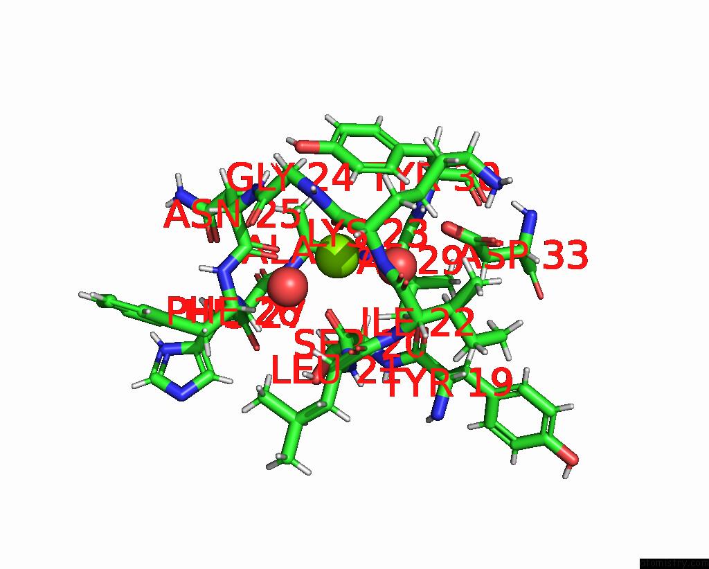
Mono view
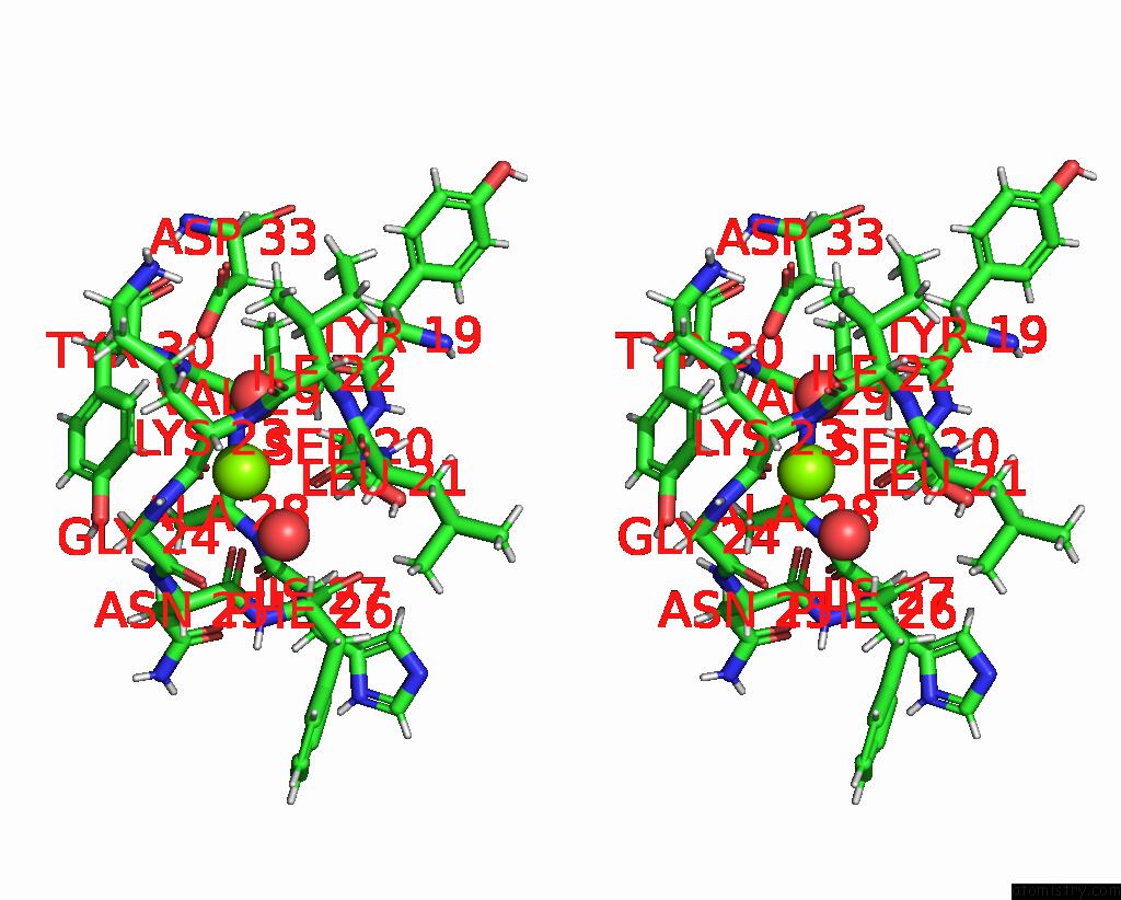
Stereo pair view
Mono view
Stereo pair view
A full contact list of Magnesium with other atoms in the Mg binding
site number 1 of Crystal Structure of ZN2+ Bound Calprotectin within 5.0Å range:
|
Magnesium binding site 2 out of 2 in 8sjc
Go back to
Magnesium binding site 2 out
of 2 in the Crystal Structure of ZN2+ Bound Calprotectin
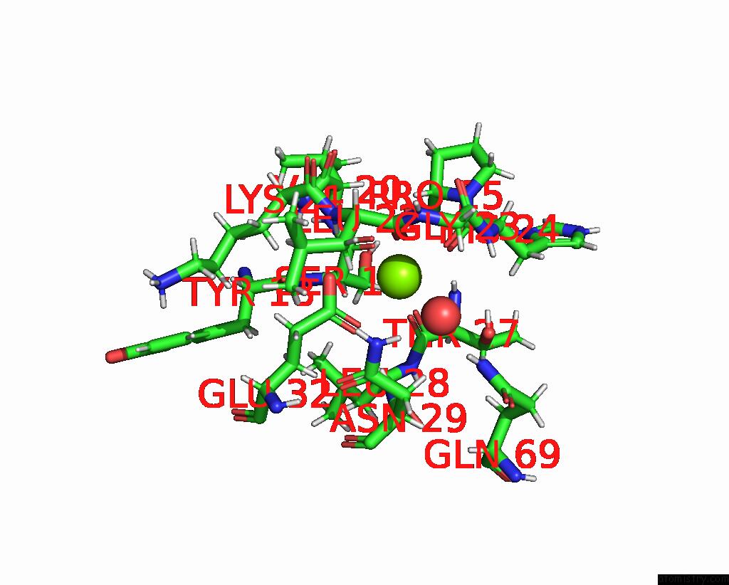
Mono view
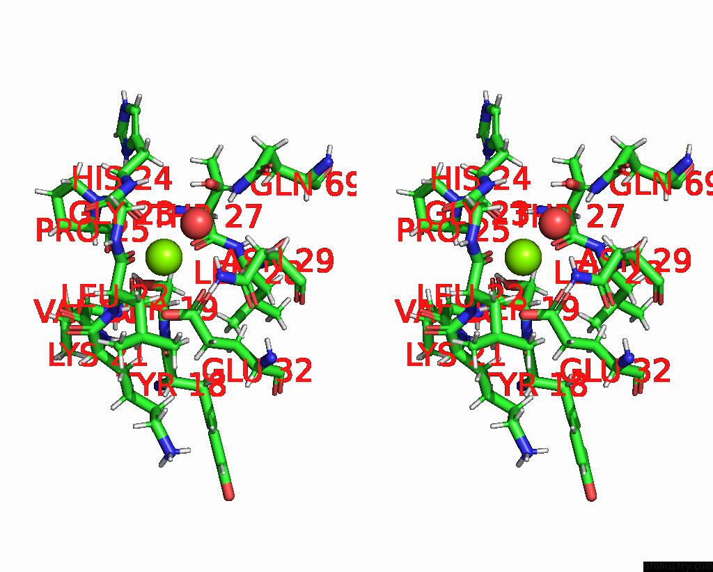
Stereo pair view
Mono view
Stereo pair view
A full contact list of Magnesium with other atoms in the Mg binding
site number 2 of Crystal Structure of ZN2+ Bound Calprotectin within 5.0Å range:
|
Reference:
Y.R.Perera,
V.Garcia,
R.M.Guillen,
W.J.Chazin.
Crystal Structure of ZN2+ Bound Calprotectin To Be Published.
Page generated: Fri Aug 15 15:47:19 2025
Last articles
Mn in 1CNZMn in 1CKN
Mn in 1CJT
Mn in 1CJK
Mn in 1CE8
Mn in 1CEV
Mn in 1BFR
Mn in 1C3O
Mn in 1CDK
Mn in 1C30