Magnesium »
PDB 8wcl-8woo »
8wdg »
Magnesium in PDB 8wdg: Subatomic Crystal Structure of Glucose Isomerase From Streptomyces Rubiginosus
Protein crystallography data
The structure of Subatomic Crystal Structure of Glucose Isomerase From Streptomyces Rubiginosus, PDB code: 8wdg
was solved by
K.H.Nam,
with X-Ray Crystallography technique. A brief refinement statistics is given in the table below:
| Resolution Low / High (Å) | 40.77 / 0.99 |
| Space group | I 2 2 2 |
| Cell size a, b, c (Å), α, β, γ (°) | 92.675, 98.626, 102.137, 90, 90, 90 |
| R / Rfree (%) | 15.8 / 17.1 |
Magnesium Binding Sites:
The binding sites of Magnesium atom in the Subatomic Crystal Structure of Glucose Isomerase From Streptomyces Rubiginosus
(pdb code 8wdg). This binding sites where shown within
5.0 Angstroms radius around Magnesium atom.
In total 3 binding sites of Magnesium where determined in the Subatomic Crystal Structure of Glucose Isomerase From Streptomyces Rubiginosus, PDB code: 8wdg:
Jump to Magnesium binding site number: 1; 2; 3;
In total 3 binding sites of Magnesium where determined in the Subatomic Crystal Structure of Glucose Isomerase From Streptomyces Rubiginosus, PDB code: 8wdg:
Jump to Magnesium binding site number: 1; 2; 3;
Magnesium binding site 1 out of 3 in 8wdg
Go back to
Magnesium binding site 1 out
of 3 in the Subatomic Crystal Structure of Glucose Isomerase From Streptomyces Rubiginosus
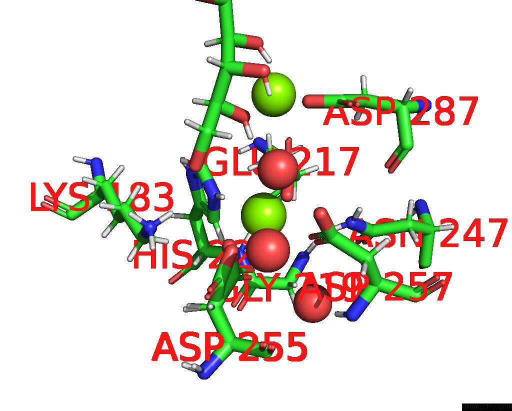
Mono view
Stereo pair view
Mono view
Stereo pair view
A full contact list of Magnesium with other atoms in the Mg binding
site number 1 of Subatomic Crystal Structure of Glucose Isomerase From Streptomyces Rubiginosus within 5.0Å range:
|
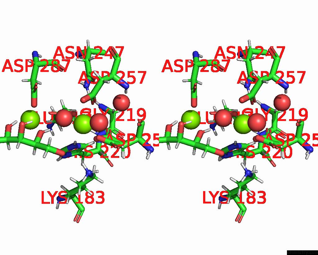
Magnesium binding site 2 out of 3 in 8wdg
Go back to
Magnesium binding site 2 out
of 3 in the Subatomic Crystal Structure of Glucose Isomerase From Streptomyces Rubiginosus

Mono view
Stereo pair view
Mono view
Stereo pair view
A full contact list of Magnesium with other atoms in the Mg binding
site number 2 of Subatomic Crystal Structure of Glucose Isomerase From Streptomyces Rubiginosus within 5.0Å range:
|
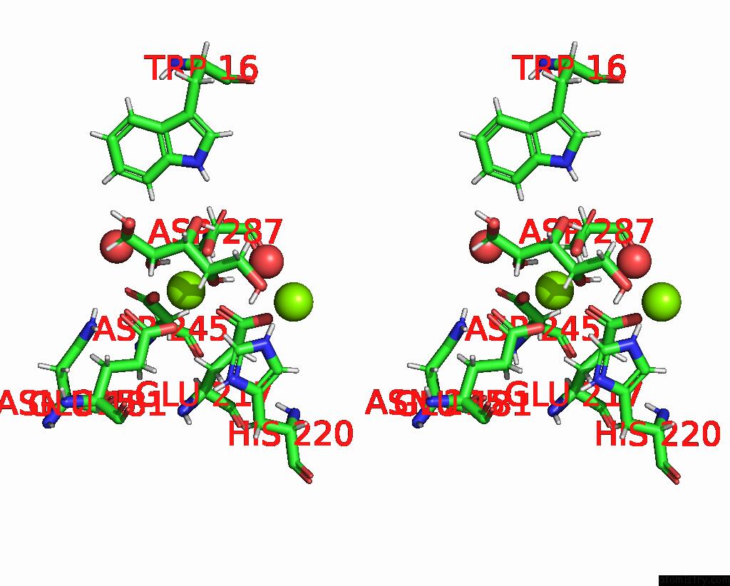
Magnesium binding site 3 out of 3 in 8wdg
Go back to
Magnesium binding site 3 out
of 3 in the Subatomic Crystal Structure of Glucose Isomerase From Streptomyces Rubiginosus

Mono view

Stereo pair view
Mono view
Stereo pair view
A full contact list of Magnesium with other atoms in the Mg binding
site number 3 of Subatomic Crystal Structure of Glucose Isomerase From Streptomyces Rubiginosus within 5.0Å range:
|
Reference:
K.H.Nam,
K.H.Nam.
N/A N/A.
Page generated: Fri Aug 15 18:43:27 2025
Last articles
Mg in 8XPAMg in 8XOY
Mg in 8XOQ
Mg in 8XOO
Mg in 8XON
Mg in 8XOM
Mg in 8XOL
Mg in 8XOC
Mg in 8XO2
Mg in 8XNE