Magnesium »
PDB 8zj7-8zwq »
8zk7 »
Magnesium in PDB 8zk7: Crystal Structure of the Decarboxylase KDC4427 Mutant E468L From Enterobacter Sp. Cgmcc 5087
Protein crystallography data
The structure of Crystal Structure of the Decarboxylase KDC4427 Mutant E468L From Enterobacter Sp. Cgmcc 5087, PDB code: 8zk7
was solved by
S.Dong,
L.Liu,
H.Zhang,
with X-Ray Crystallography technique. A brief refinement statistics is given in the table below:
| Resolution Low / High (Å) | 68.53 / 2.32 |
| Space group | P 21 21 2 |
| Cell size a, b, c (Å), α, β, γ (°) | 116.604, 137.054, 77.488, 90, 90, 90 |
| R / Rfree (%) | 20.3 / 24.7 |
Magnesium Binding Sites:
The binding sites of Magnesium atom in the Crystal Structure of the Decarboxylase KDC4427 Mutant E468L From Enterobacter Sp. Cgmcc 5087
(pdb code 8zk7). This binding sites where shown within
5.0 Angstroms radius around Magnesium atom.
In total 2 binding sites of Magnesium where determined in the Crystal Structure of the Decarboxylase KDC4427 Mutant E468L From Enterobacter Sp. Cgmcc 5087, PDB code: 8zk7:
Jump to Magnesium binding site number: 1; 2;
In total 2 binding sites of Magnesium where determined in the Crystal Structure of the Decarboxylase KDC4427 Mutant E468L From Enterobacter Sp. Cgmcc 5087, PDB code: 8zk7:
Jump to Magnesium binding site number: 1; 2;
Magnesium binding site 1 out of 2 in 8zk7
Go back to
Magnesium binding site 1 out
of 2 in the Crystal Structure of the Decarboxylase KDC4427 Mutant E468L From Enterobacter Sp. Cgmcc 5087
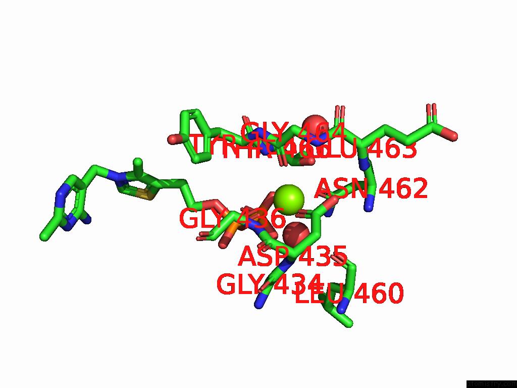
Mono view
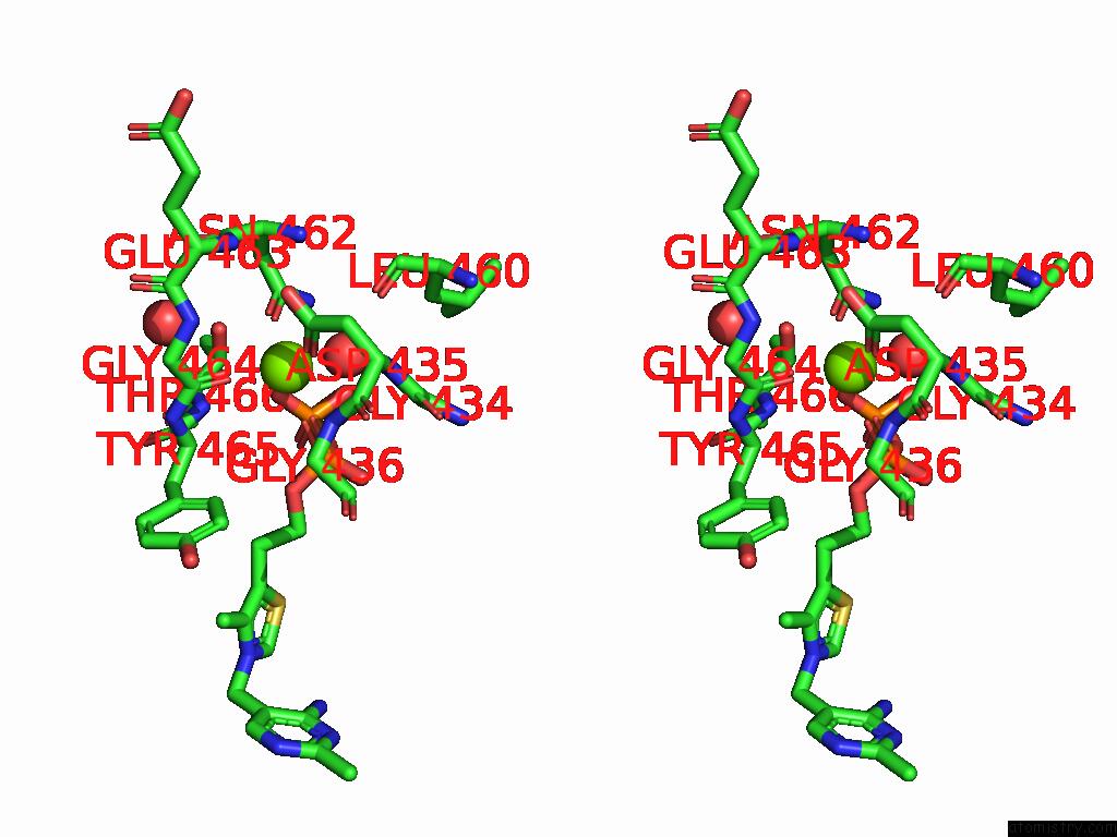
Stereo pair view
Mono view
Stereo pair view
A full contact list of Magnesium with other atoms in the Mg binding
site number 1 of Crystal Structure of the Decarboxylase KDC4427 Mutant E468L From Enterobacter Sp. Cgmcc 5087 within 5.0Å range:
|
Magnesium binding site 2 out of 2 in 8zk7
Go back to
Magnesium binding site 2 out
of 2 in the Crystal Structure of the Decarboxylase KDC4427 Mutant E468L From Enterobacter Sp. Cgmcc 5087
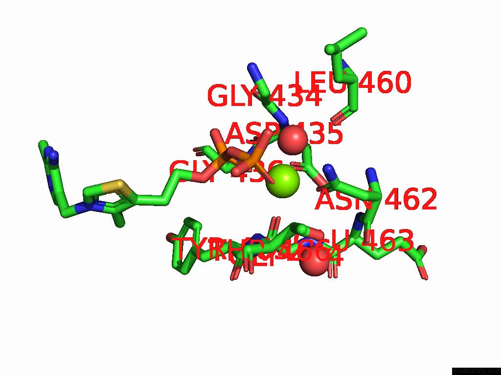
Mono view
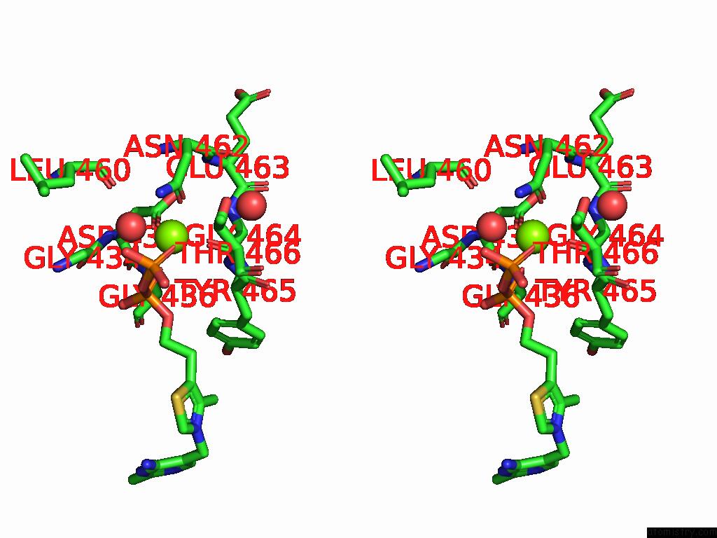
Stereo pair view
Mono view
Stereo pair view
A full contact list of Magnesium with other atoms in the Mg binding
site number 2 of Crystal Structure of the Decarboxylase KDC4427 Mutant E468L From Enterobacter Sp. Cgmcc 5087 within 5.0Å range:
|
Reference:
S.Dong,
L.Liu,
H.Zhang.
Crystal Structure of the Decarboxylase KDC4427 Mutant E468L From Enterobacter Sp. Cgmcc 5087 To Be Published.
Page generated: Fri Aug 15 22:41:51 2025
Last articles
Mn in 5O5LMn in 5O5K
Mn in 5O7F
Mn in 5O6N
Mn in 5O6I
Mn in 5O6G
Mn in 5NTD
Mn in 5O3N
Mn in 5O25
Mn in 5NSM