Magnesium »
PDB 8zj7-8zwq »
8zk9 »
Magnesium in PDB 8zk9: Crystal Structure of the Decarboxylase KDC4427 Mutant E468L in Complex with Phenylpyruvic Acid
Protein crystallography data
The structure of Crystal Structure of the Decarboxylase KDC4427 Mutant E468L in Complex with Phenylpyruvic Acid, PDB code: 8zk9
was solved by
S.Dong,
L.Liu,
H.Zhang,
with X-Ray Crystallography technique. A brief refinement statistics is given in the table below:
| Resolution Low / High (Å) | 64.27 / 2.03 |
| Space group | P 21 21 2 |
| Cell size a, b, c (Å), α, β, γ (°) | 115.916, 136.52, 77.234, 90, 90, 90 |
| R / Rfree (%) | 22 / 25.4 |
Magnesium Binding Sites:
The binding sites of Magnesium atom in the Crystal Structure of the Decarboxylase KDC4427 Mutant E468L in Complex with Phenylpyruvic Acid
(pdb code 8zk9). This binding sites where shown within
5.0 Angstroms radius around Magnesium atom.
In total 2 binding sites of Magnesium where determined in the Crystal Structure of the Decarboxylase KDC4427 Mutant E468L in Complex with Phenylpyruvic Acid, PDB code: 8zk9:
Jump to Magnesium binding site number: 1; 2;
In total 2 binding sites of Magnesium where determined in the Crystal Structure of the Decarboxylase KDC4427 Mutant E468L in Complex with Phenylpyruvic Acid, PDB code: 8zk9:
Jump to Magnesium binding site number: 1; 2;
Magnesium binding site 1 out of 2 in 8zk9
Go back to
Magnesium binding site 1 out
of 2 in the Crystal Structure of the Decarboxylase KDC4427 Mutant E468L in Complex with Phenylpyruvic Acid
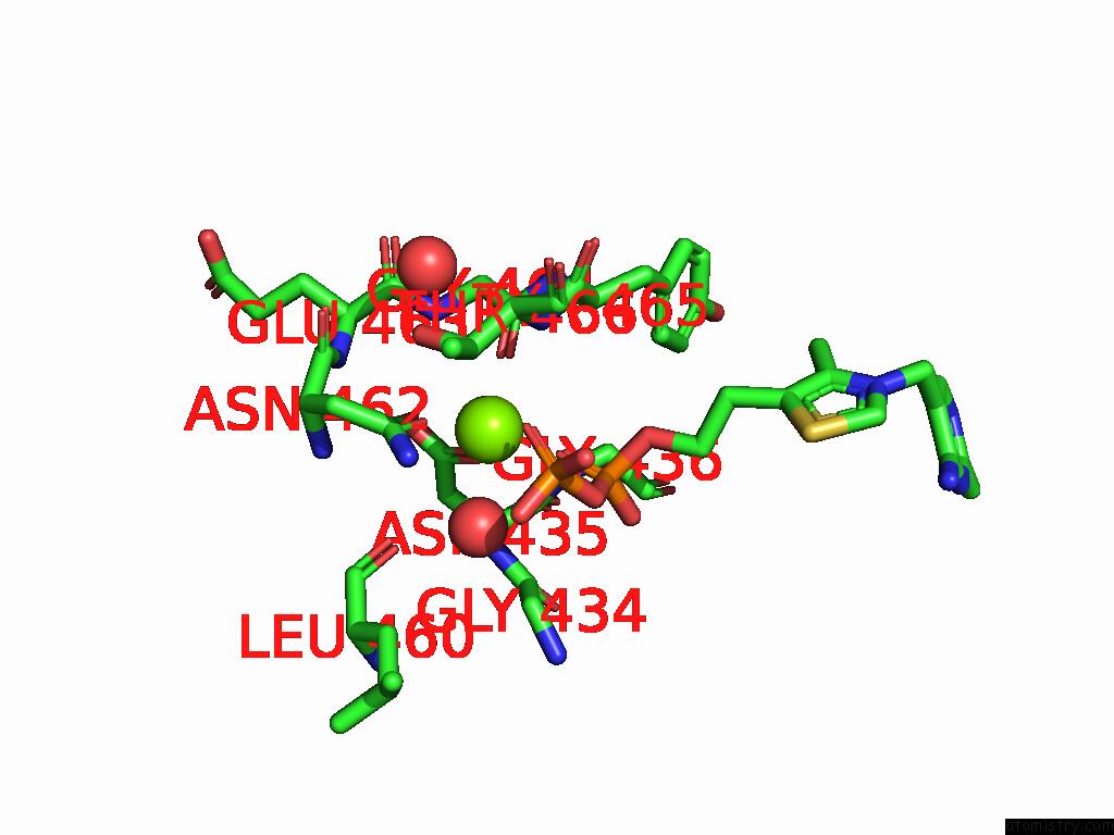
Mono view
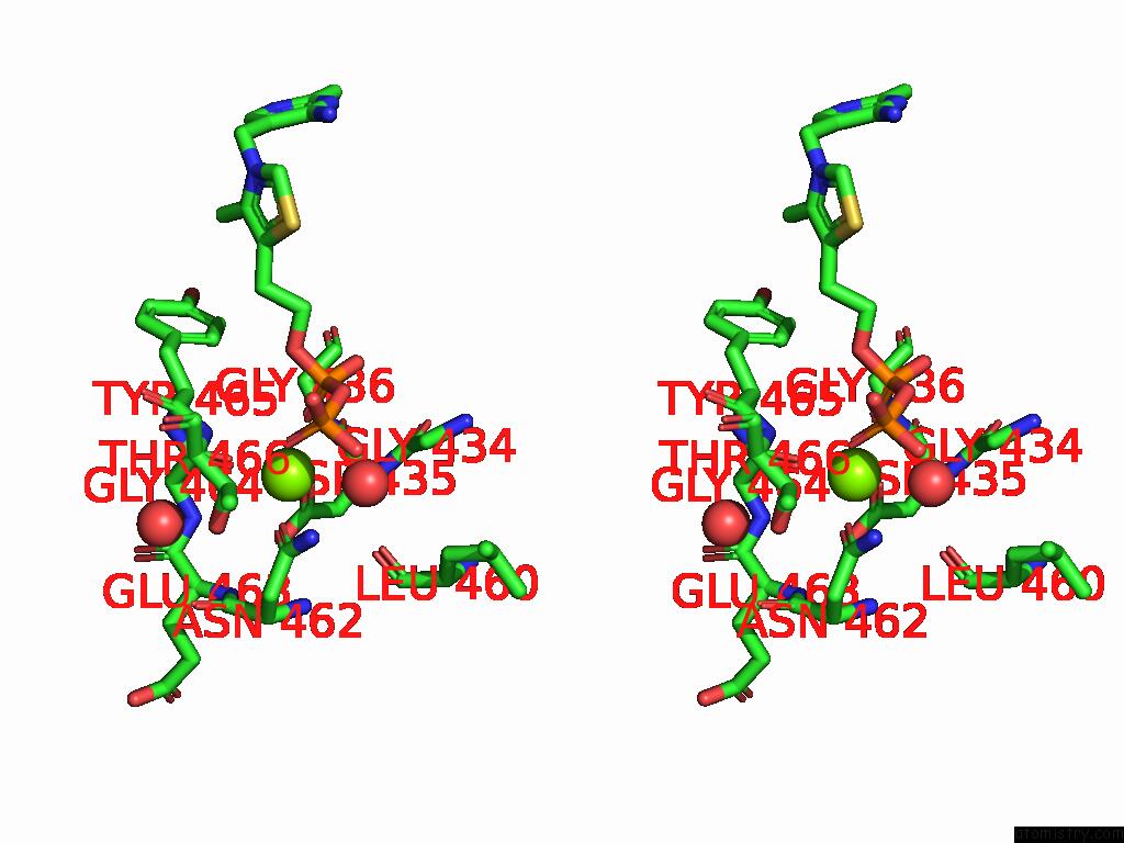
Stereo pair view
Mono view
Stereo pair view
A full contact list of Magnesium with other atoms in the Mg binding
site number 1 of Crystal Structure of the Decarboxylase KDC4427 Mutant E468L in Complex with Phenylpyruvic Acid within 5.0Å range:
|
Magnesium binding site 2 out of 2 in 8zk9
Go back to
Magnesium binding site 2 out
of 2 in the Crystal Structure of the Decarboxylase KDC4427 Mutant E468L in Complex with Phenylpyruvic Acid
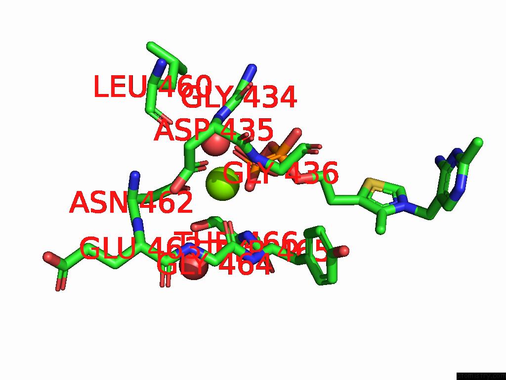
Mono view
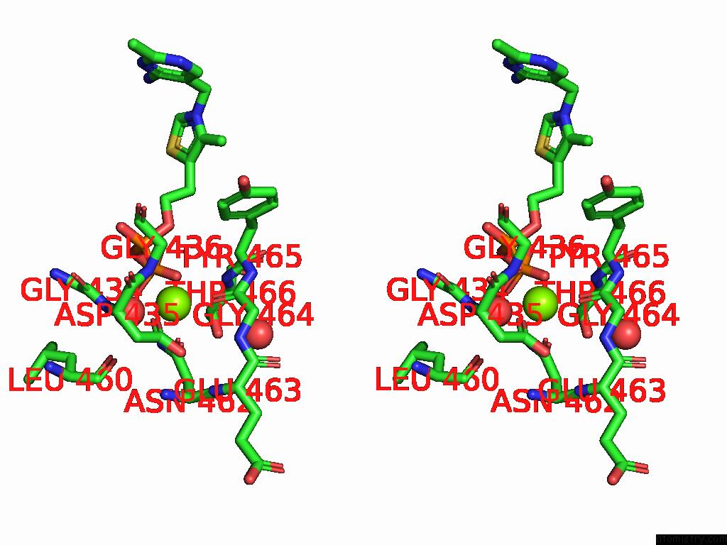
Stereo pair view
Mono view
Stereo pair view
A full contact list of Magnesium with other atoms in the Mg binding
site number 2 of Crystal Structure of the Decarboxylase KDC4427 Mutant E468L in Complex with Phenylpyruvic Acid within 5.0Å range:
|
Reference:
S.Dong,
L.Liu,
H.Zhang.
Crystal Structure of the Decarboxylase KDC4427 Mutant E468L in Complex with Indole-3-Pyruvic Acid To Be Published.
Page generated: Fri Aug 15 22:42:23 2025
Last articles
Mn in 4WIUMn in 4WIE
Mn in 4WFO
Mn in 4WFA
Mn in 4UXA
Mn in 4W8Y
Mn in 4W9S
Mn in 4V15
Mn in 4V0U
Mn in 4V0W