Magnesium »
PDB 8zj7-8zwq »
8zn7 »
Magnesium in PDB 8zn7: Crystal Structure of Designed Clock Protein Kaic
Protein crystallography data
The structure of Crystal Structure of Designed Clock Protein Kaic, PDB code: 8zn7
was solved by
Y.Furuike,
S.Akiyama,
with X-Ray Crystallography technique. A brief refinement statistics is given in the table below:
| Resolution Low / High (Å) | 49.19 / 2.54 |
| Space group | P 1 21 1 |
| Cell size a, b, c (Å), α, β, γ (°) | 91.977, 175.183, 94.774, 90, 96.93, 90 |
| R / Rfree (%) | 22.5 / 27.2 |
Magnesium Binding Sites:
Pages:
>>> Page 1 <<< Page 2, Binding sites: 11 - 12;Binding sites:
The binding sites of Magnesium atom in the Crystal Structure of Designed Clock Protein Kaic (pdb code 8zn7). This binding sites where shown within 5.0 Angstroms radius around Magnesium atom.In total 12 binding sites of Magnesium where determined in the Crystal Structure of Designed Clock Protein Kaic, PDB code: 8zn7:
Jump to Magnesium binding site number: 1; 2; 3; 4; 5; 6; 7; 8; 9; 10;
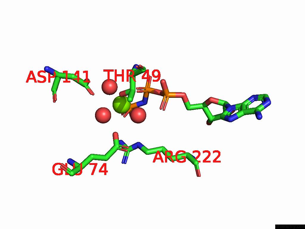
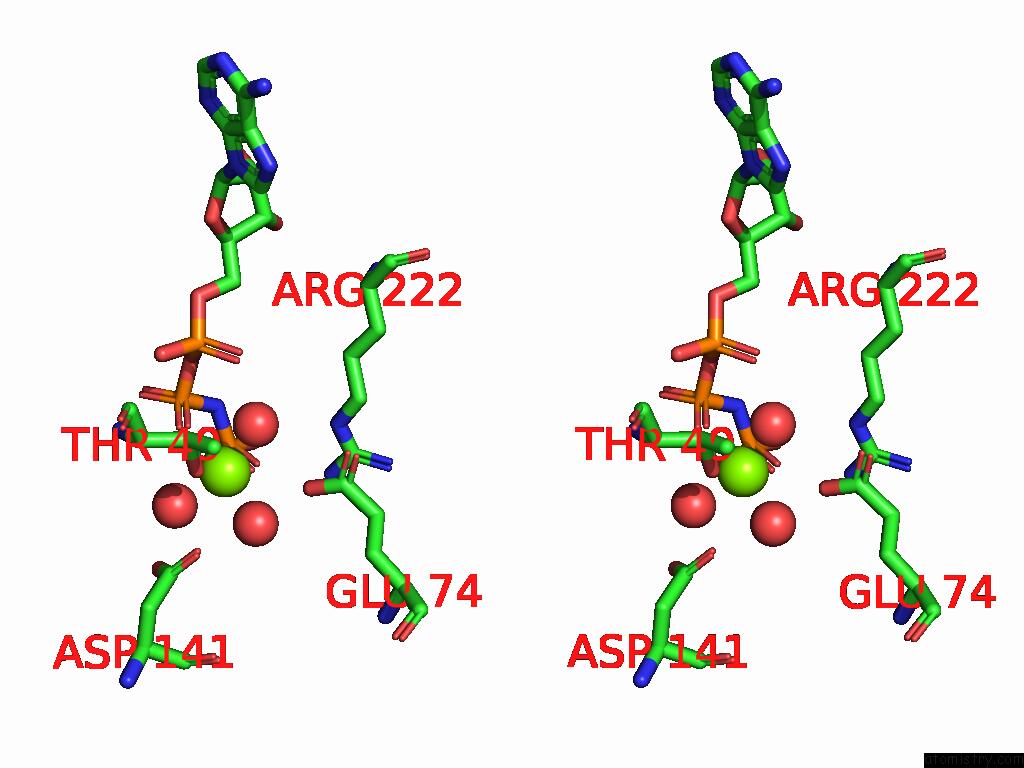
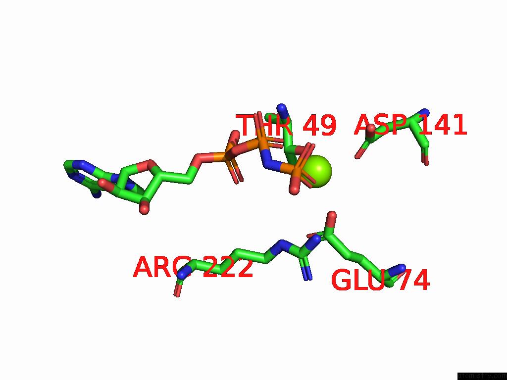
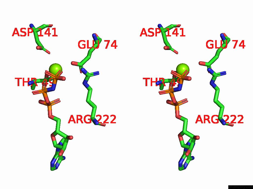
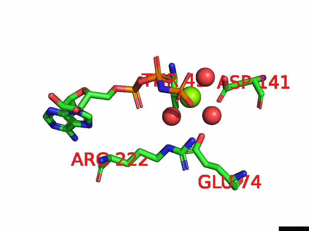
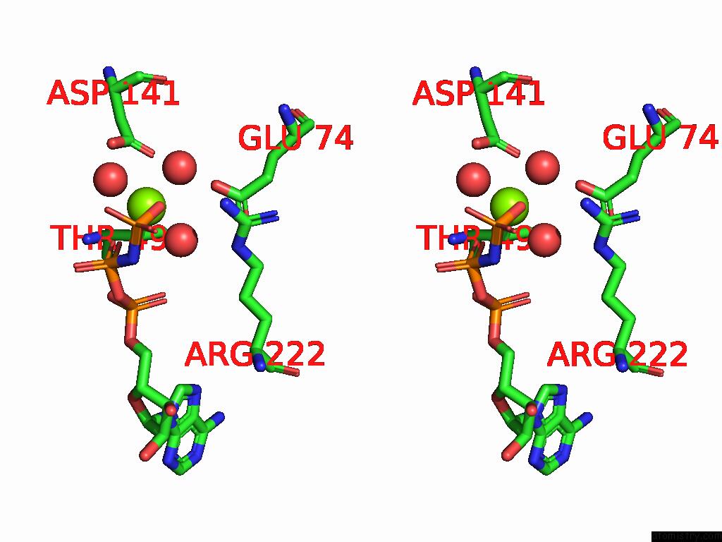
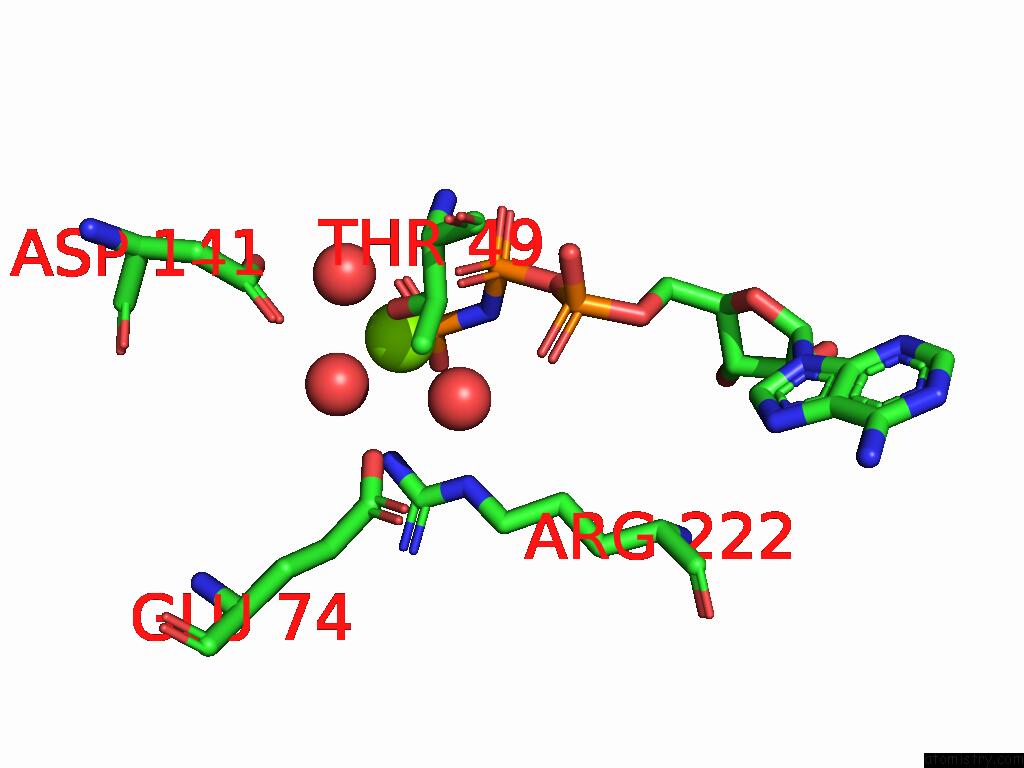
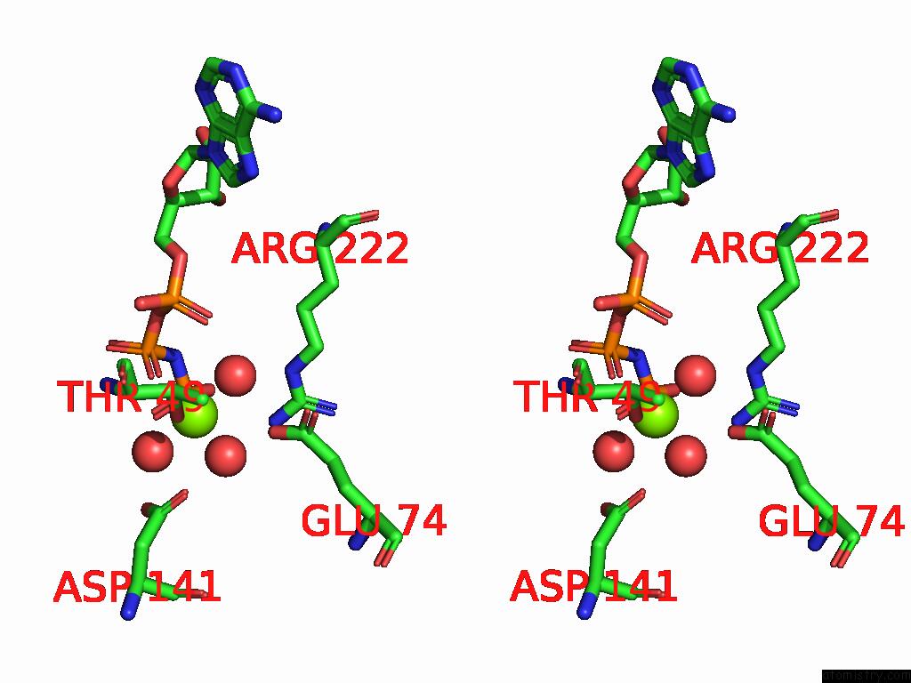
Magnesium binding site 1 out of 12 in 8zn7
Go back to
Magnesium binding site 1 out
of 12 in the Crystal Structure of Designed Clock Protein Kaic
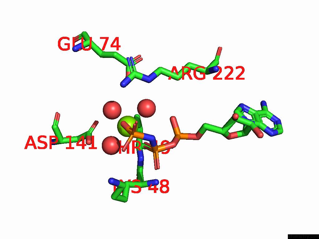
Mono view
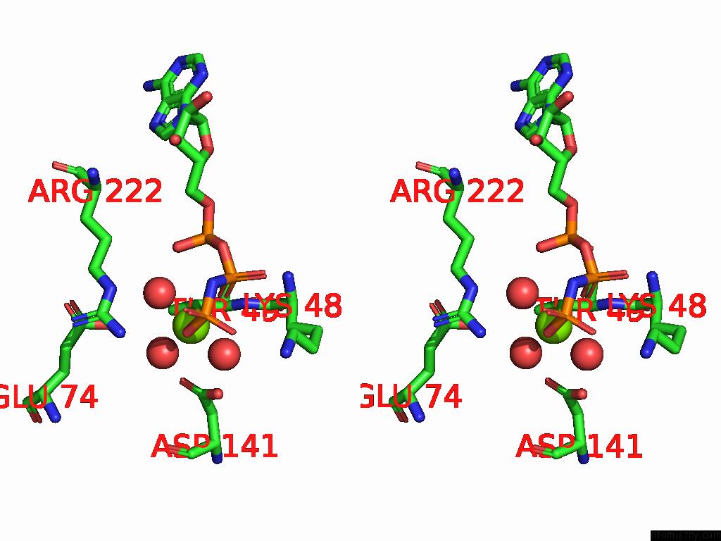
Stereo pair view
Mono view
Stereo pair view
A full contact list of Magnesium with other atoms in the Mg binding
site number 1 of Crystal Structure of Designed Clock Protein Kaic within 5.0Å range:
|
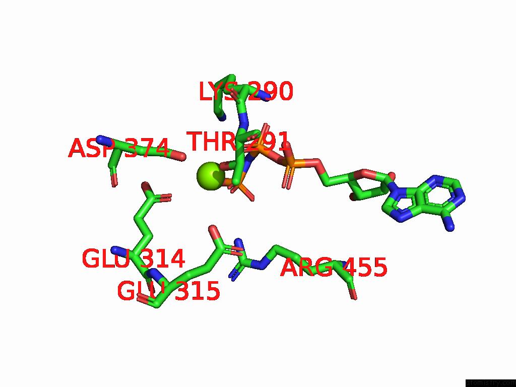
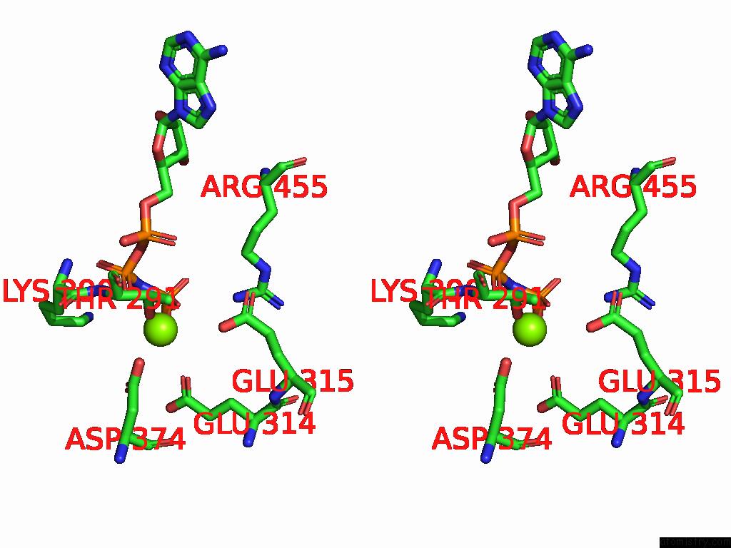
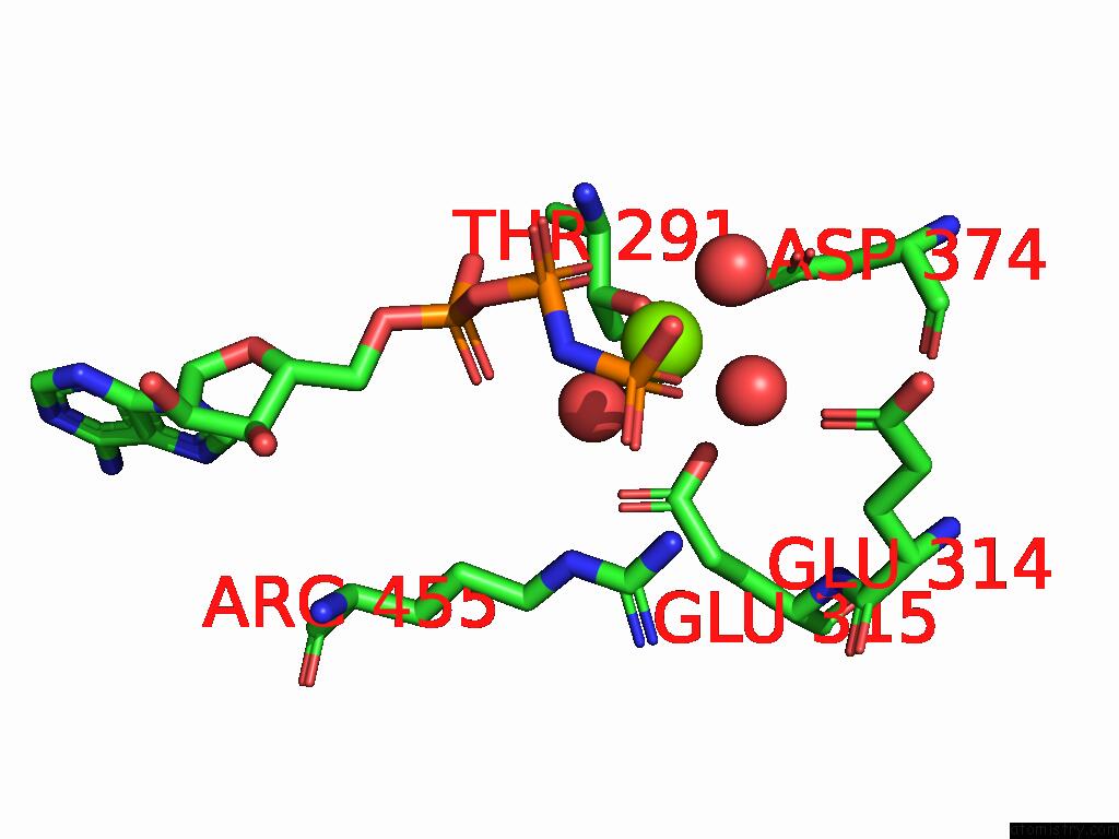
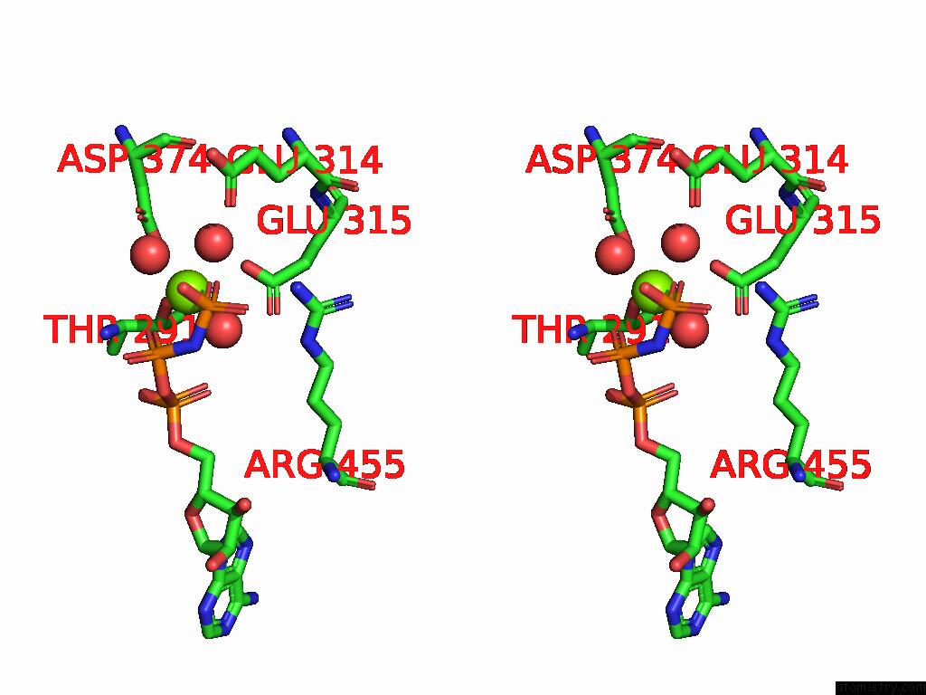
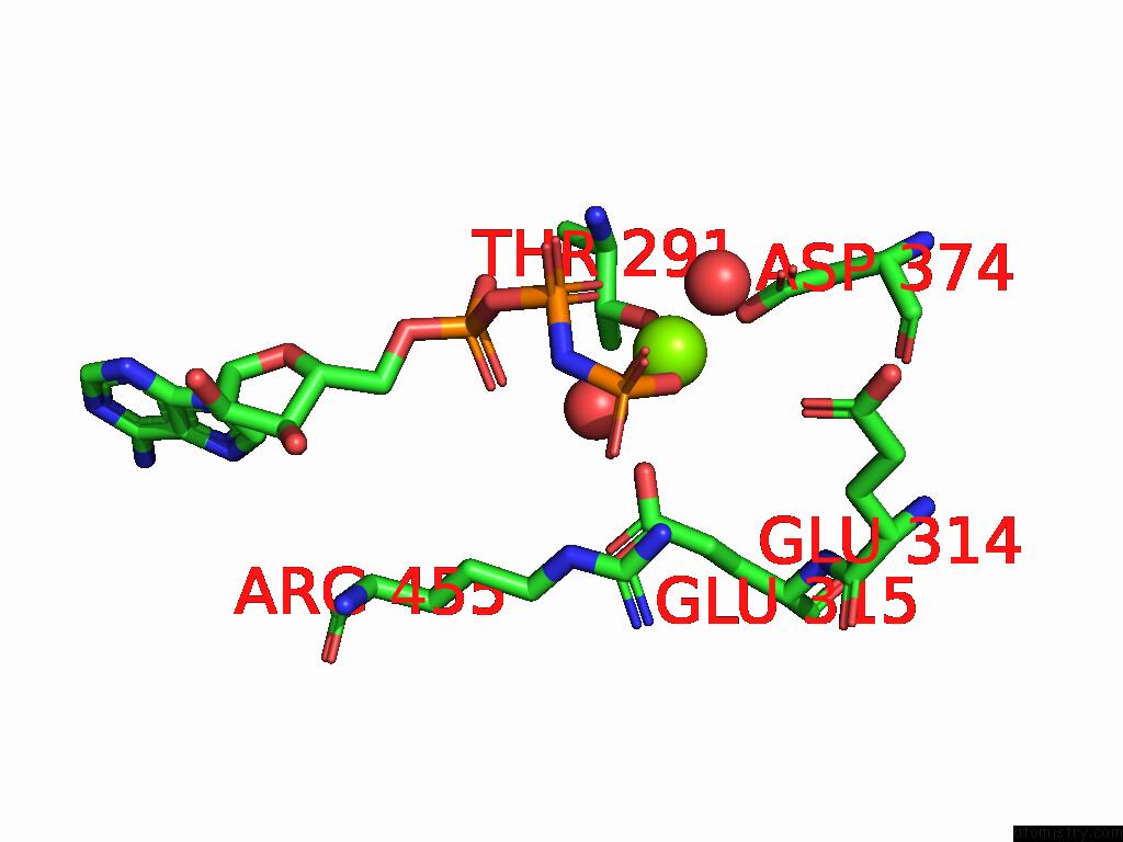
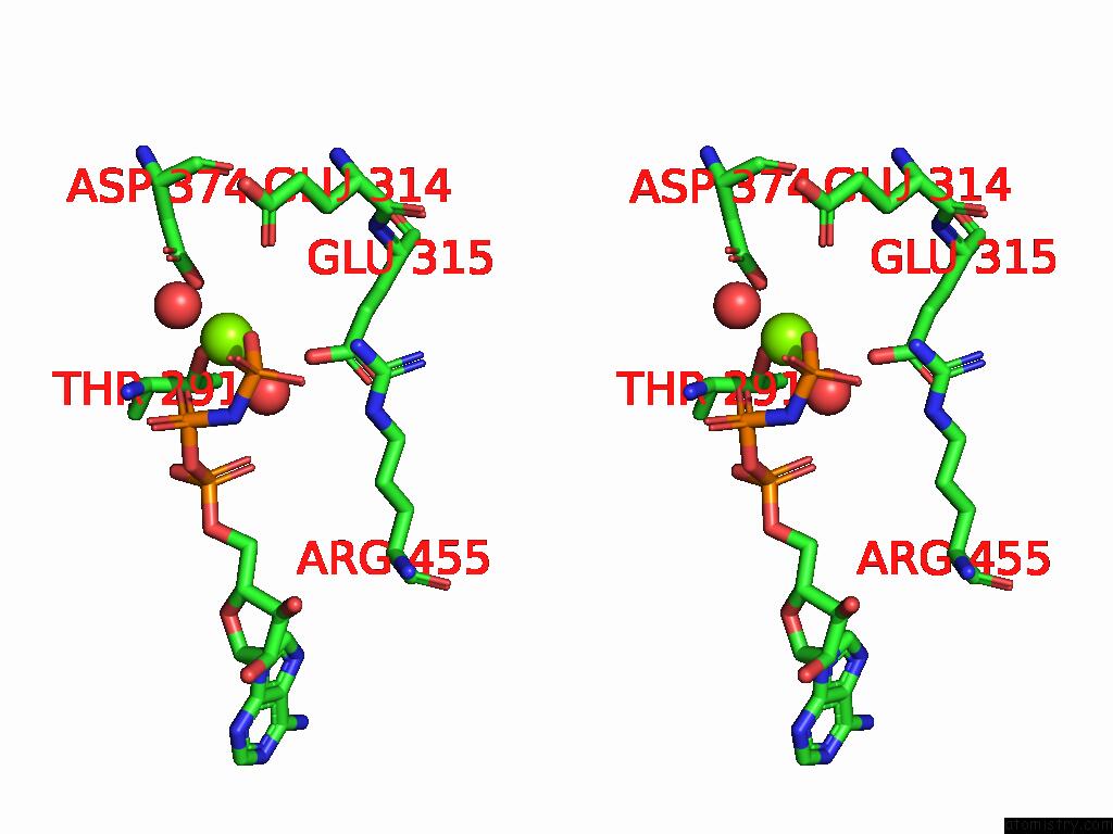
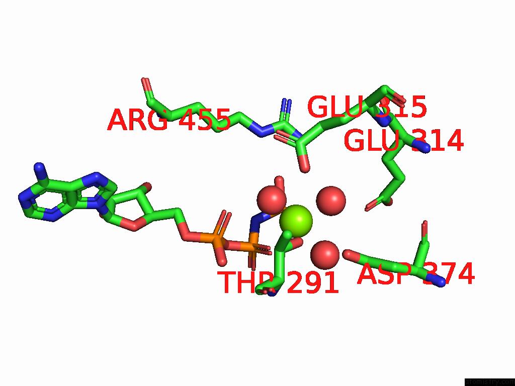
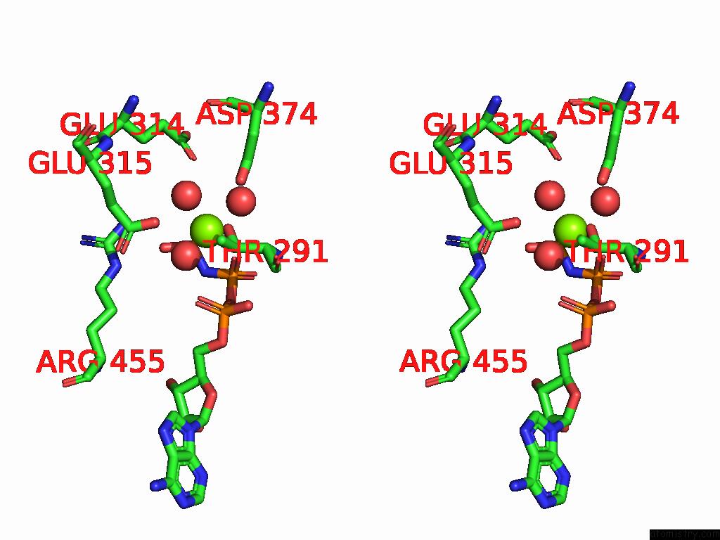
Magnesium binding site 2 out of 12 in 8zn7
Go back to
Magnesium binding site 2 out
of 12 in the Crystal Structure of Designed Clock Protein Kaic
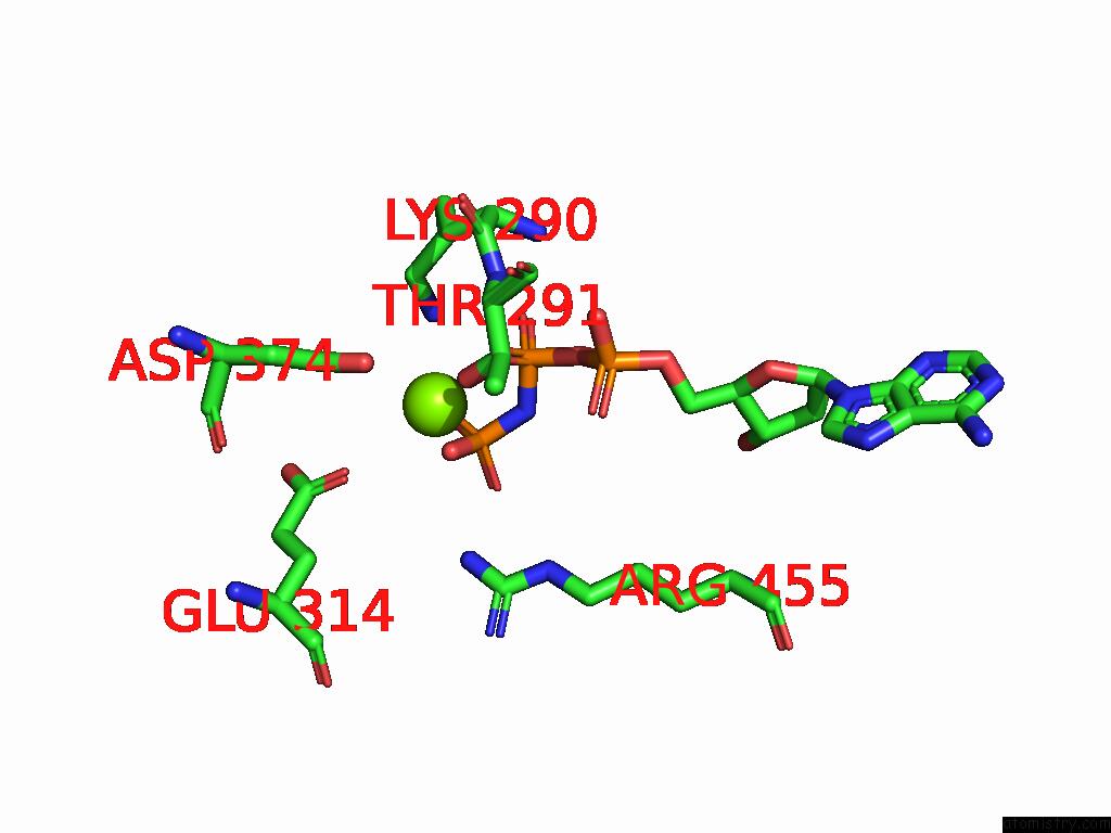
Mono view
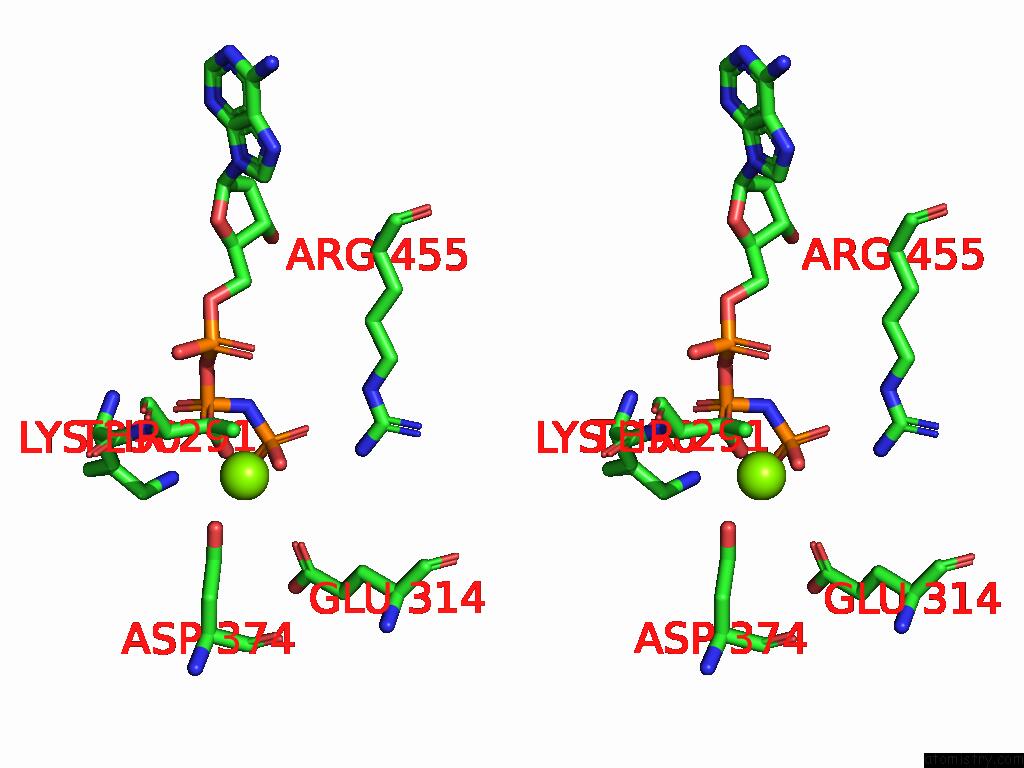
Stereo pair view
Mono view
Stereo pair view
A full contact list of Magnesium with other atoms in the Mg binding
site number 2 of Crystal Structure of Designed Clock Protein Kaic within 5.0Å range:
|
Magnesium binding site 3 out of 12 in 8zn7
Go back to
Magnesium binding site 3 out
of 12 in the Crystal Structure of Designed Clock Protein Kaic
Mono view
Stereo pair view
Mono view
Stereo pair view
A full contact list of Magnesium with other atoms in the Mg binding
site number 3 of Crystal Structure of Designed Clock Protein Kaic within 5.0Å range:
|
Magnesium binding site 4 out of 12 in 8zn7
Go back to
Magnesium binding site 4 out
of 12 in the Crystal Structure of Designed Clock Protein Kaic
Mono view
Stereo pair view
Mono view
Stereo pair view
A full contact list of Magnesium with other atoms in the Mg binding
site number 4 of Crystal Structure of Designed Clock Protein Kaic within 5.0Å range:
|
Magnesium binding site 5 out of 12 in 8zn7
Go back to
Magnesium binding site 5 out
of 12 in the Crystal Structure of Designed Clock Protein Kaic
Mono view
Stereo pair view
Mono view
Stereo pair view
A full contact list of Magnesium with other atoms in the Mg binding
site number 5 of Crystal Structure of Designed Clock Protein Kaic within 5.0Å range:
|
Magnesium binding site 6 out of 12 in 8zn7
Go back to
Magnesium binding site 6 out
of 12 in the Crystal Structure of Designed Clock Protein Kaic
Mono view
Stereo pair view
Mono view
Stereo pair view
A full contact list of Magnesium with other atoms in the Mg binding
site number 6 of Crystal Structure of Designed Clock Protein Kaic within 5.0Å range:
|
Magnesium binding site 7 out of 12 in 8zn7
Go back to
Magnesium binding site 7 out
of 12 in the Crystal Structure of Designed Clock Protein Kaic
Mono view
Stereo pair view
Mono view
Stereo pair view
A full contact list of Magnesium with other atoms in the Mg binding
site number 7 of Crystal Structure of Designed Clock Protein Kaic within 5.0Å range:
|
Magnesium binding site 8 out of 12 in 8zn7
Go back to
Magnesium binding site 8 out
of 12 in the Crystal Structure of Designed Clock Protein Kaic
Mono view
Stereo pair view
Mono view
Stereo pair view
A full contact list of Magnesium with other atoms in the Mg binding
site number 8 of Crystal Structure of Designed Clock Protein Kaic within 5.0Å range:
|
Magnesium binding site 9 out of 12 in 8zn7
Go back to
Magnesium binding site 9 out
of 12 in the Crystal Structure of Designed Clock Protein Kaic
Mono view
Stereo pair view
Mono view
Stereo pair view
A full contact list of Magnesium with other atoms in the Mg binding
site number 9 of Crystal Structure of Designed Clock Protein Kaic within 5.0Å range:
|
Magnesium binding site 10 out of 12 in 8zn7
Go back to
Magnesium binding site 10 out
of 12 in the Crystal Structure of Designed Clock Protein Kaic
Mono view
Stereo pair view
Mono view
Stereo pair view
A full contact list of Magnesium with other atoms in the Mg binding
site number 10 of Crystal Structure of Designed Clock Protein Kaic within 5.0Å range:
|
Reference:
Y.Furuike,
S.Akiyama.
Crystal Structure of Designed Clock Protein Kaic To Be Published.
Page generated: Fri Aug 15 22:48:39 2025
Last articles
Mn in 6H2PMn in 6H21
Mn in 6H2M
Mn in 6H1M
Mn in 6H0B
Mn in 6H08
Mn in 6GXC
Mn in 6H07
Mn in 6GYX
Mn in 6GIU