Magnesium »
PDB 8zxg-9b27 »
8zxg »
Magnesium in PDB 8zxg: Crystal Structure of Paraoxonase From Bacillus Sp. Strain S3WAHI
Protein crystallography data
The structure of Crystal Structure of Paraoxonase From Bacillus Sp. Strain S3WAHI, PDB code: 8zxg
was solved by
A.A.Azman,
N.D.Muhd Noor,
A.T.C.Leow,
S.A.Mohd Noor,
M.S.Mohamad Ali,
with X-Ray Crystallography technique. A brief refinement statistics is given in the table below:
| Resolution Low / High (Å) | 39.01 / 1.49 |
| Space group | P 41 21 2 |
| Cell size a, b, c (Å), α, β, γ (°) | 77.823, 77.823, 135.266, 90, 90, 90 |
| R / Rfree (%) | 19.3 / n/a |
Other elements in 8zxg:
The structure of Crystal Structure of Paraoxonase From Bacillus Sp. Strain S3WAHI also contains other interesting chemical elements:
| Zinc | (Zn) | 2 atoms |
Magnesium Binding Sites:
Pages:
>>> Page 1 <<< Page 2, Binding sites: 11 - 20; Page 3, Binding sites: 21 - 30; Page 4, Binding sites: 31 - 40; Page 5, Binding sites: 41 - 50; Page 6, Binding sites: 51 - 58;Binding sites:
The binding sites of Magnesium atom in the Crystal Structure of Paraoxonase From Bacillus Sp. Strain S3WAHI (pdb code 8zxg). This binding sites where shown within 5.0 Angstroms radius around Magnesium atom.In total 58 binding sites of Magnesium where determined in the Crystal Structure of Paraoxonase From Bacillus Sp. Strain S3WAHI, PDB code: 8zxg:
Jump to Magnesium binding site number: 1; 2; 3; 4; 5; 6; 7; 8; 9; 10;
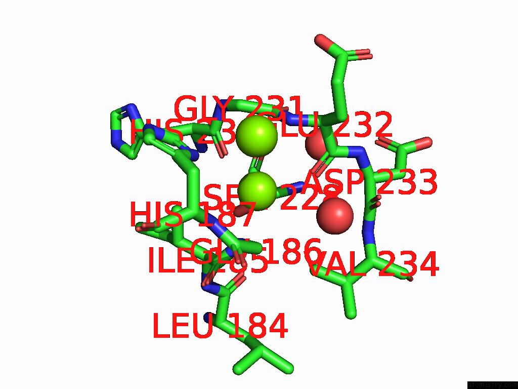
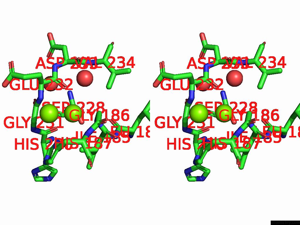
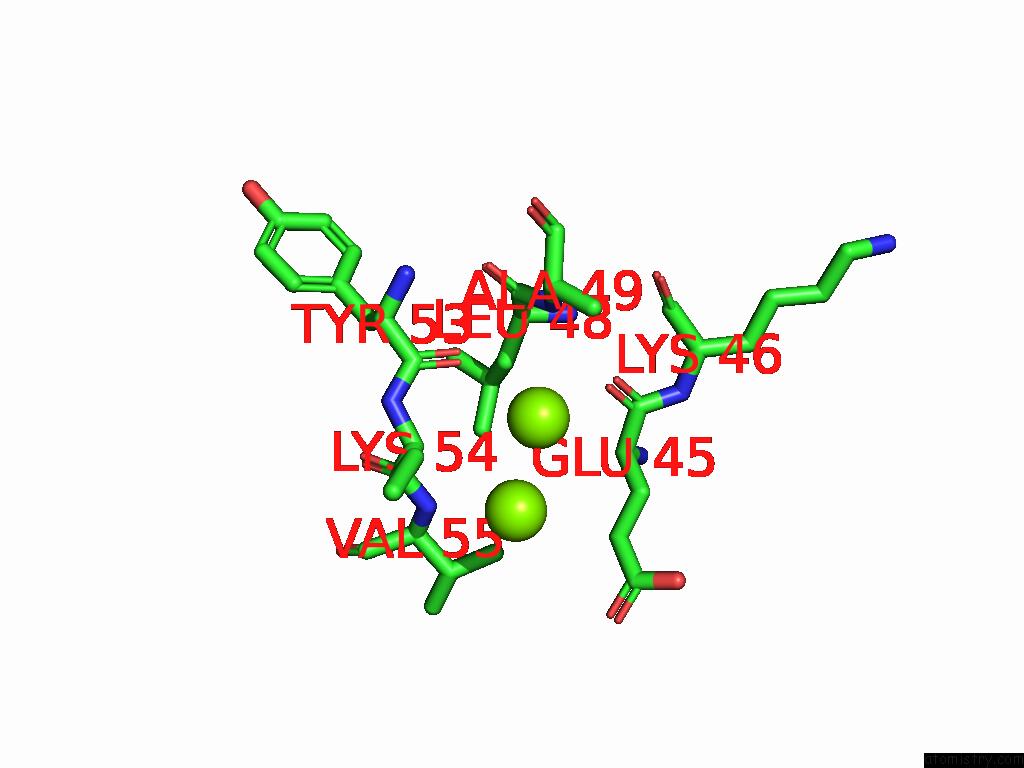
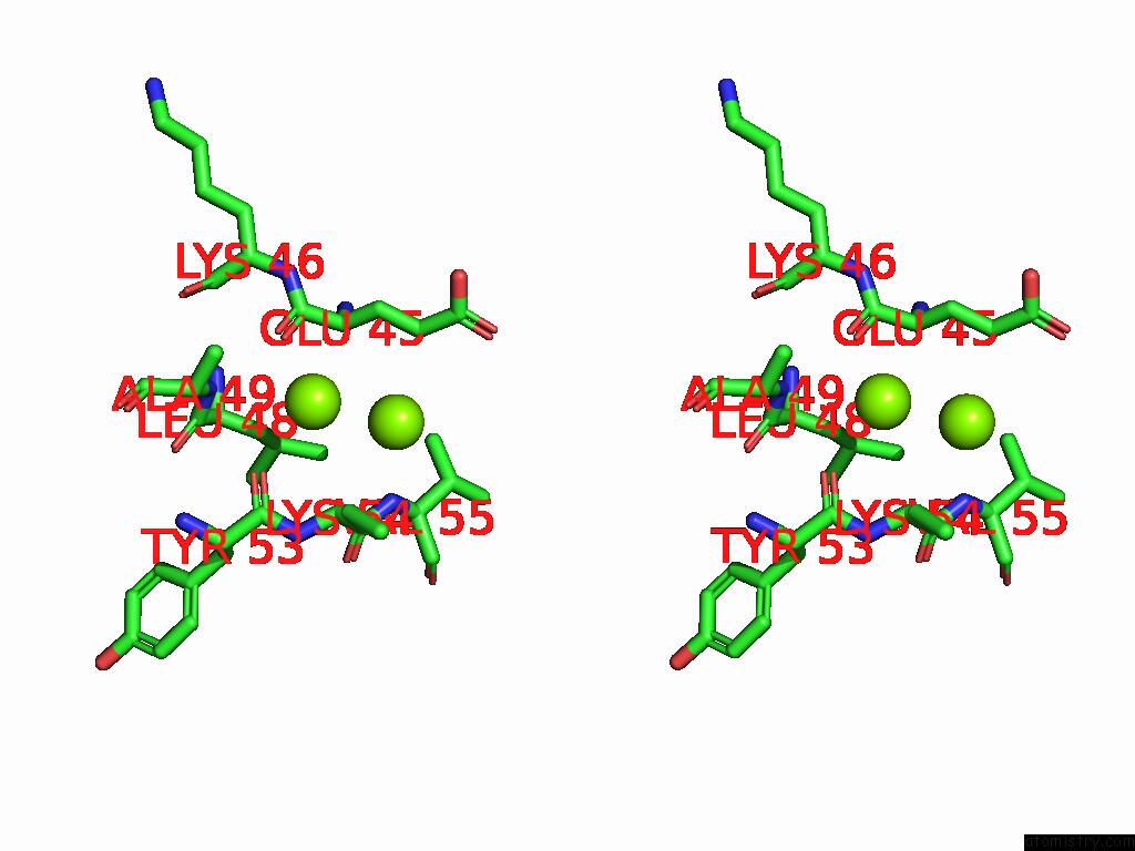
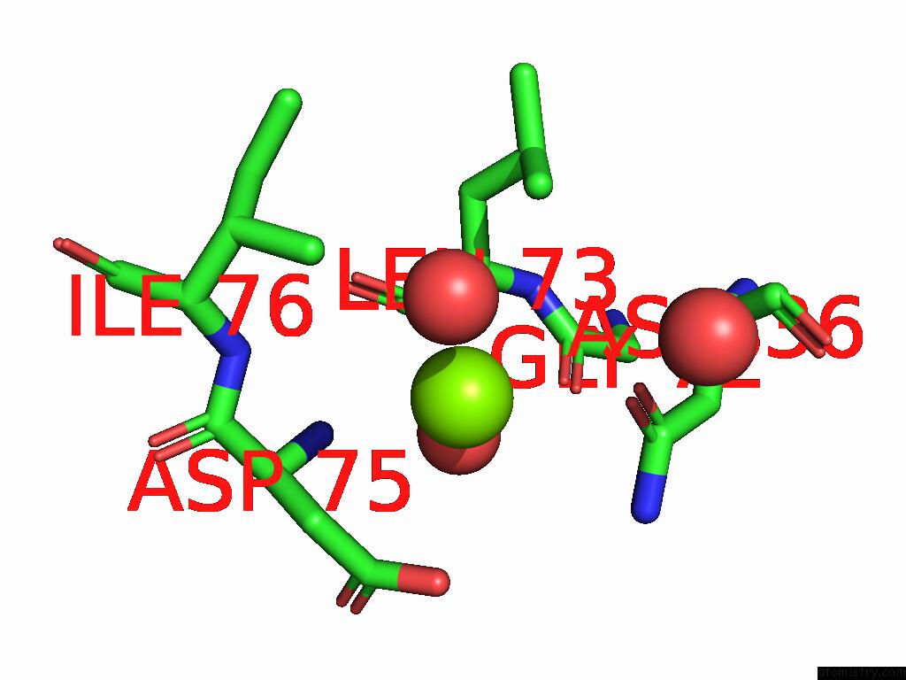
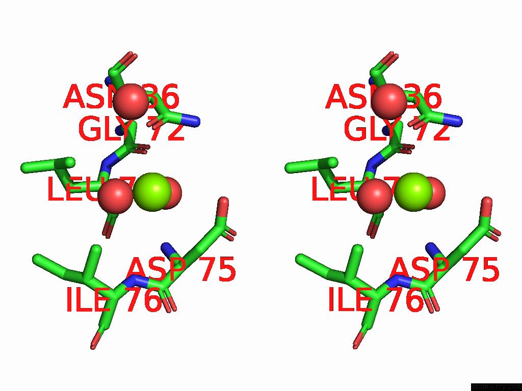
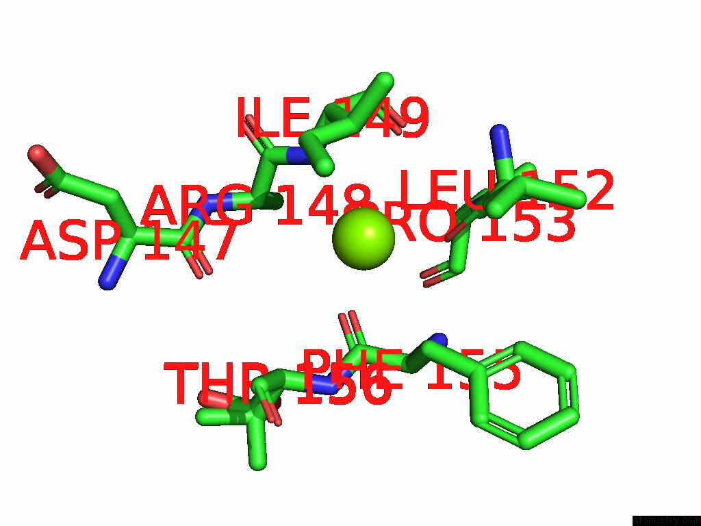
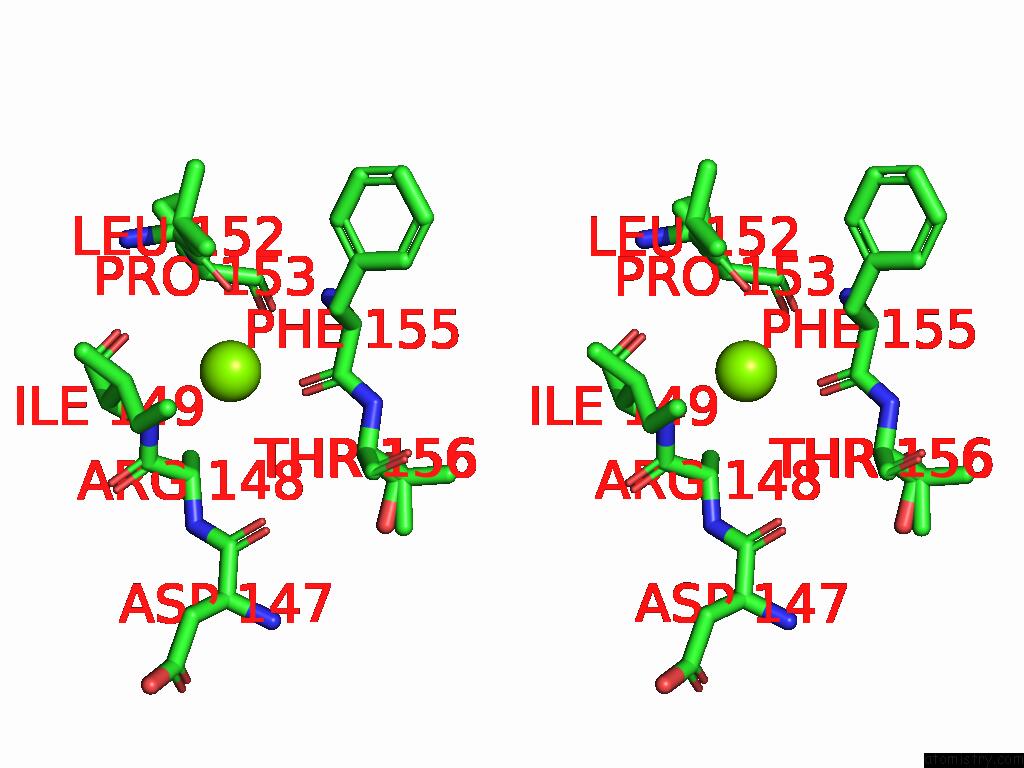
Magnesium binding site 1 out of 58 in 8zxg
Go back to
Magnesium binding site 1 out
of 58 in the Crystal Structure of Paraoxonase From Bacillus Sp. Strain S3WAHI
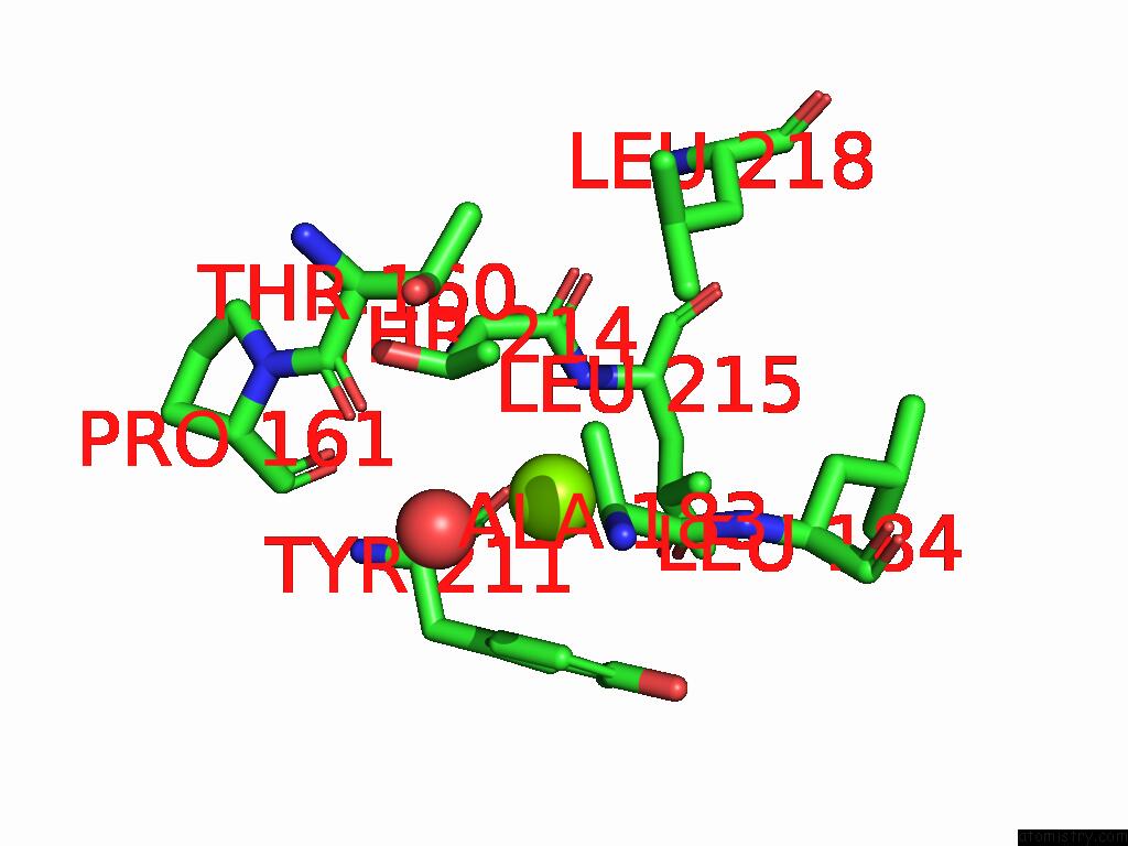
Mono view
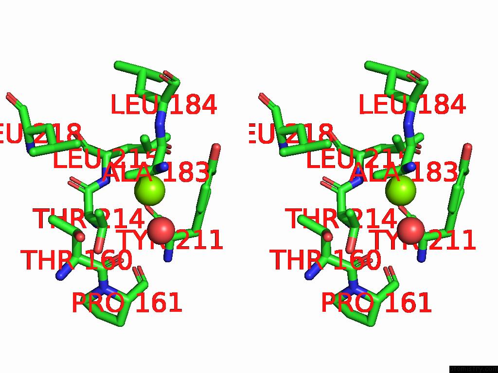
Stereo pair view
Mono view
Stereo pair view
A full contact list of Magnesium with other atoms in the Mg binding
site number 1 of Crystal Structure of Paraoxonase From Bacillus Sp. Strain S3WAHI within 5.0Å range:
|
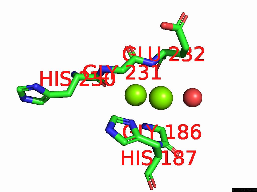
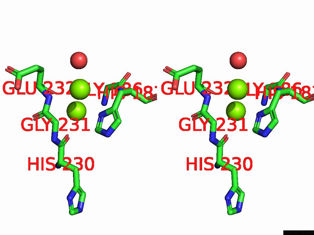
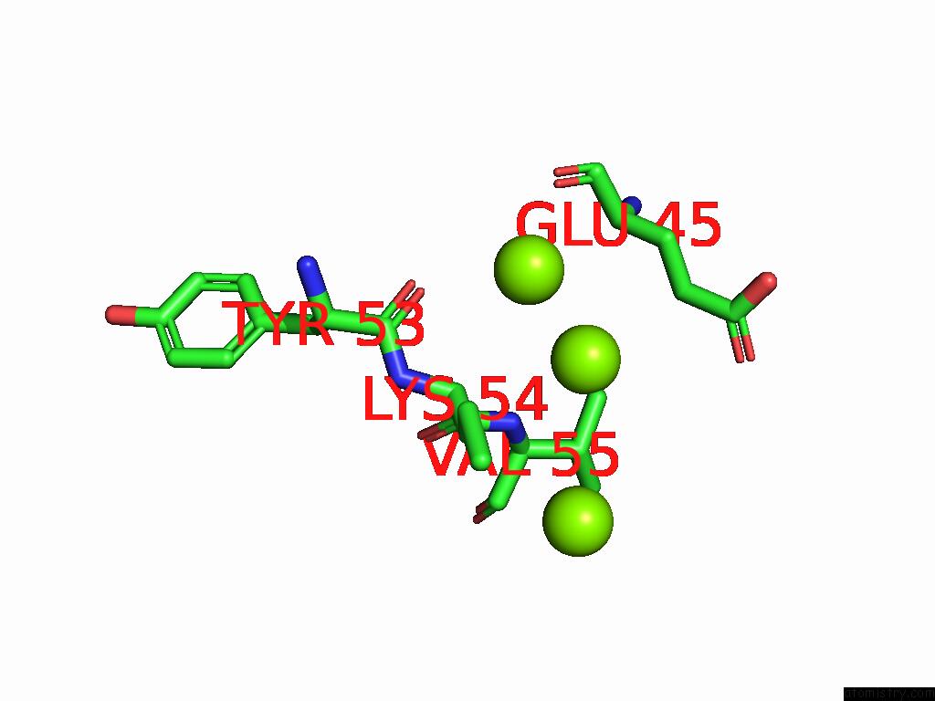
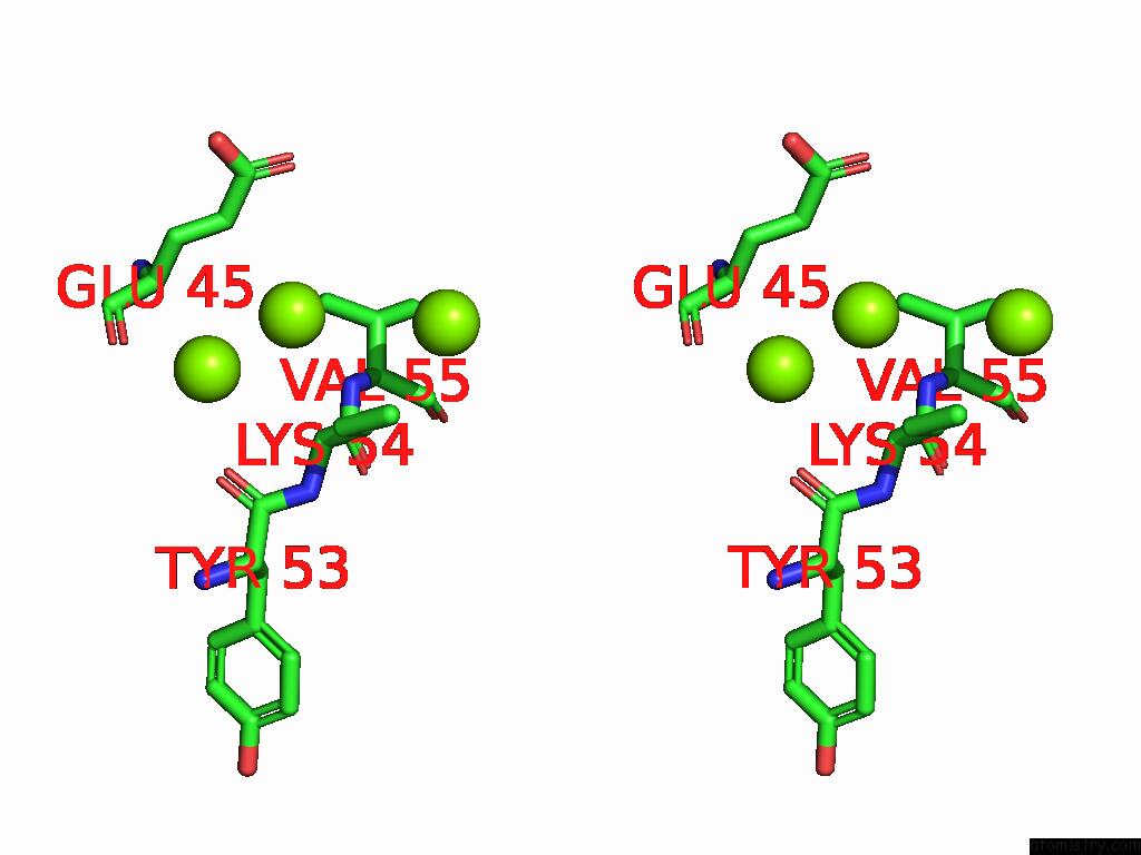
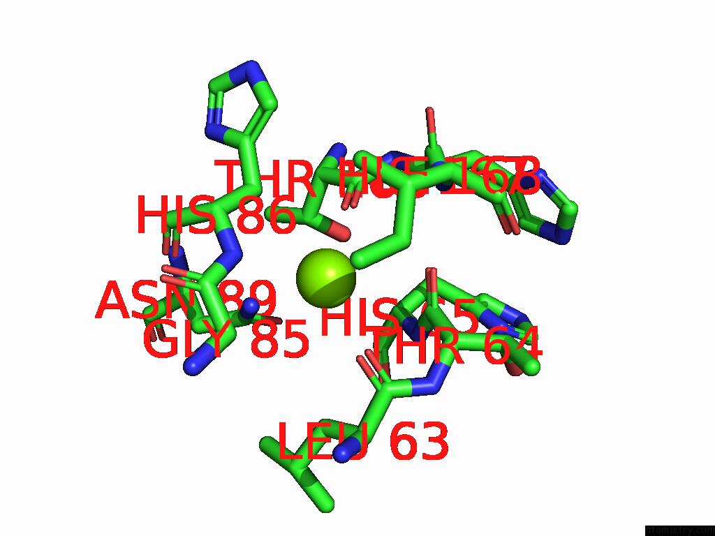
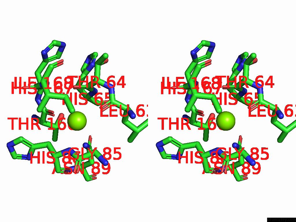
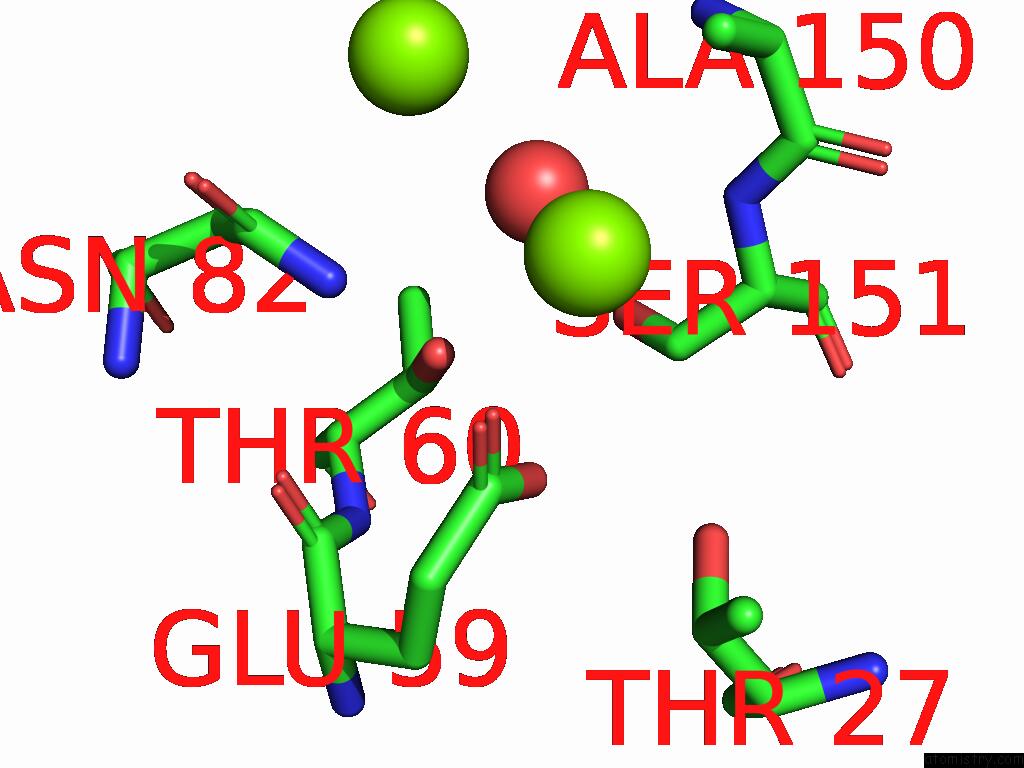
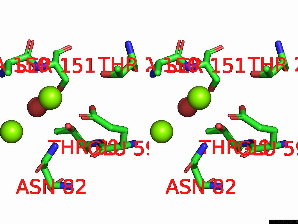
Magnesium binding site 2 out of 58 in 8zxg
Go back to
Magnesium binding site 2 out
of 58 in the Crystal Structure of Paraoxonase From Bacillus Sp. Strain S3WAHI
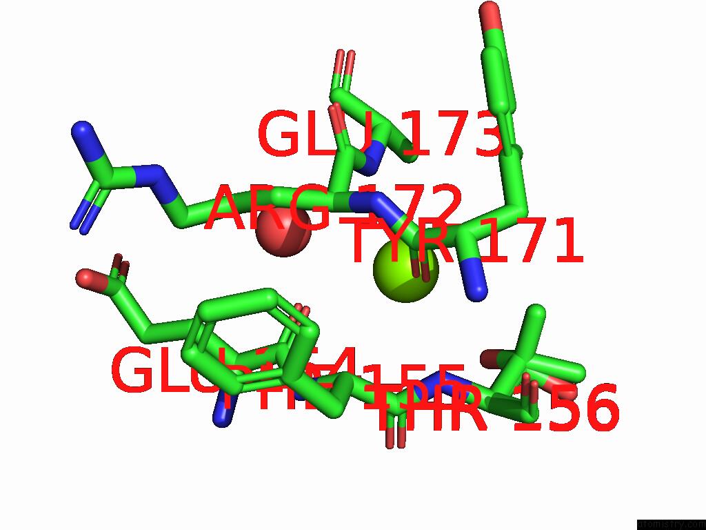
Mono view
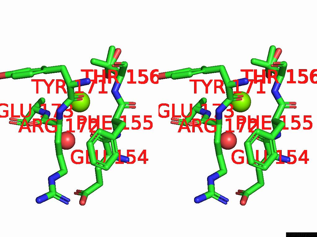
Stereo pair view
Mono view
Stereo pair view
A full contact list of Magnesium with other atoms in the Mg binding
site number 2 of Crystal Structure of Paraoxonase From Bacillus Sp. Strain S3WAHI within 5.0Å range:
|
Magnesium binding site 3 out of 58 in 8zxg
Go back to
Magnesium binding site 3 out
of 58 in the Crystal Structure of Paraoxonase From Bacillus Sp. Strain S3WAHI
Mono view
Stereo pair view
Mono view
Stereo pair view
A full contact list of Magnesium with other atoms in the Mg binding
site number 3 of Crystal Structure of Paraoxonase From Bacillus Sp. Strain S3WAHI within 5.0Å range:
|
Magnesium binding site 4 out of 58 in 8zxg
Go back to
Magnesium binding site 4 out
of 58 in the Crystal Structure of Paraoxonase From Bacillus Sp. Strain S3WAHI
Mono view
Stereo pair view
Mono view
Stereo pair view
A full contact list of Magnesium with other atoms in the Mg binding
site number 4 of Crystal Structure of Paraoxonase From Bacillus Sp. Strain S3WAHI within 5.0Å range:
|
Magnesium binding site 5 out of 58 in 8zxg
Go back to
Magnesium binding site 5 out
of 58 in the Crystal Structure of Paraoxonase From Bacillus Sp. Strain S3WAHI
Mono view
Stereo pair view
Mono view
Stereo pair view
A full contact list of Magnesium with other atoms in the Mg binding
site number 5 of Crystal Structure of Paraoxonase From Bacillus Sp. Strain S3WAHI within 5.0Å range:
|
Magnesium binding site 6 out of 58 in 8zxg
Go back to
Magnesium binding site 6 out
of 58 in the Crystal Structure of Paraoxonase From Bacillus Sp. Strain S3WAHI
Mono view
Stereo pair view
Mono view
Stereo pair view
A full contact list of Magnesium with other atoms in the Mg binding
site number 6 of Crystal Structure of Paraoxonase From Bacillus Sp. Strain S3WAHI within 5.0Å range:
|
Magnesium binding site 7 out of 58 in 8zxg
Go back to
Magnesium binding site 7 out
of 58 in the Crystal Structure of Paraoxonase From Bacillus Sp. Strain S3WAHI
Mono view
Stereo pair view
Mono view
Stereo pair view
A full contact list of Magnesium with other atoms in the Mg binding
site number 7 of Crystal Structure of Paraoxonase From Bacillus Sp. Strain S3WAHI within 5.0Å range:
|
Magnesium binding site 8 out of 58 in 8zxg
Go back to
Magnesium binding site 8 out
of 58 in the Crystal Structure of Paraoxonase From Bacillus Sp. Strain S3WAHI
Mono view
Stereo pair view
Mono view
Stereo pair view
A full contact list of Magnesium with other atoms in the Mg binding
site number 8 of Crystal Structure of Paraoxonase From Bacillus Sp. Strain S3WAHI within 5.0Å range:
|
Magnesium binding site 9 out of 58 in 8zxg
Go back to
Magnesium binding site 9 out
of 58 in the Crystal Structure of Paraoxonase From Bacillus Sp. Strain S3WAHI
Mono view
Stereo pair view
Mono view
Stereo pair view
A full contact list of Magnesium with other atoms in the Mg binding
site number 9 of Crystal Structure of Paraoxonase From Bacillus Sp. Strain S3WAHI within 5.0Å range:
|
Magnesium binding site 10 out of 58 in 8zxg
Go back to
Magnesium binding site 10 out
of 58 in the Crystal Structure of Paraoxonase From Bacillus Sp. Strain S3WAHI
Mono view
Stereo pair view
Mono view
Stereo pair view
A full contact list of Magnesium with other atoms in the Mg binding
site number 10 of Crystal Structure of Paraoxonase From Bacillus Sp. Strain S3WAHI within 5.0Å range:
|
Reference:
A.A.Azman,
N.D.Muhd Noor,
A.T.C.Leow,
S.A.Mohd Noor,
M.S.Mohamad Ali.
Crystal Structure of Paraoxonase From Bacillus Sp. Strain S3WAHI To Be Published.
Page generated: Fri Aug 15 23:02:15 2025
Last articles
Mn in 2QF2Mn in 2QGI
Mn in 2QEY
Mn in 2QF1
Mn in 2QCS
Mn in 2PYO
Mn in 2QEW
Mn in 2QB0
Mn in 2QB5
Mn in 2QB6