Magnesium »
PDB 9b2d-9bm0 »
9bfa »
Magnesium in PDB 9bfa: Bcat Mutant
Enzymatic activity of Bcat Mutant
All present enzymatic activity of Bcat Mutant:
2.6.1.42;
2.6.1.42;
Protein crystallography data
The structure of Bcat Mutant, PDB code: 9bfa
was solved by
M.Dong,
with X-Ray Crystallography technique. A brief refinement statistics is given in the table below:
| Resolution Low / High (Å) | 91.45 / 1.79 |
| Space group | P 21 21 21 |
| Cell size a, b, c (Å), α, β, γ (°) | 110.284, 115.63, 149.413, 90, 90, 90 |
| R / Rfree (%) | 23.2 / 26.8 |
Magnesium Binding Sites:
The binding sites of Magnesium atom in the Bcat Mutant
(pdb code 9bfa). This binding sites where shown within
5.0 Angstroms radius around Magnesium atom.
In total 3 binding sites of Magnesium where determined in the Bcat Mutant, PDB code: 9bfa:
Jump to Magnesium binding site number: 1; 2; 3;
In total 3 binding sites of Magnesium where determined in the Bcat Mutant, PDB code: 9bfa:
Jump to Magnesium binding site number: 1; 2; 3;
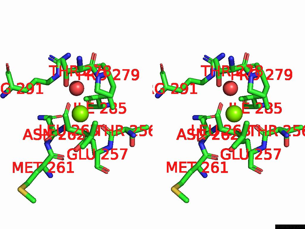
Magnesium binding site 1 out of 3 in 9bfa
Go back to
Magnesium binding site 1 out
of 3 in the Bcat Mutant

Mono view
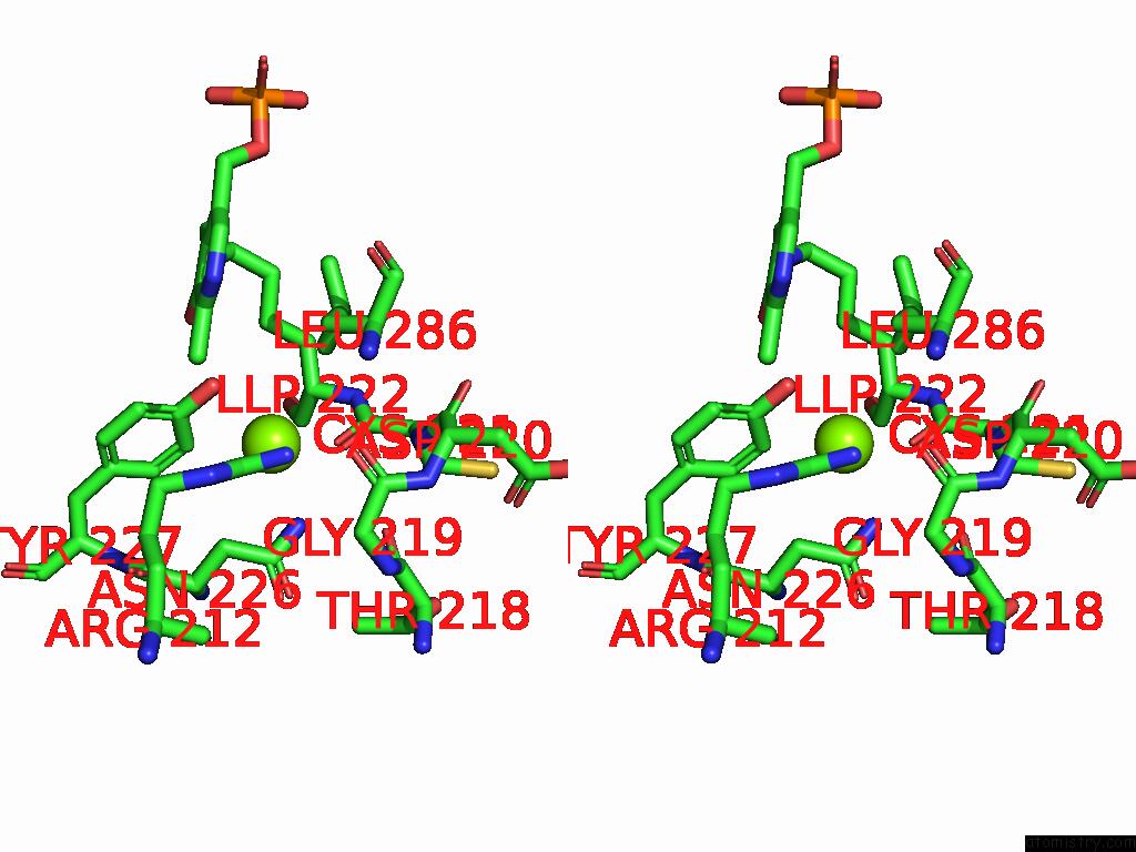
Stereo pair view
Mono view
Stereo pair view
A full contact list of Magnesium with other atoms in the Mg binding
site number 1 of Bcat Mutant within 5.0Å range:
|
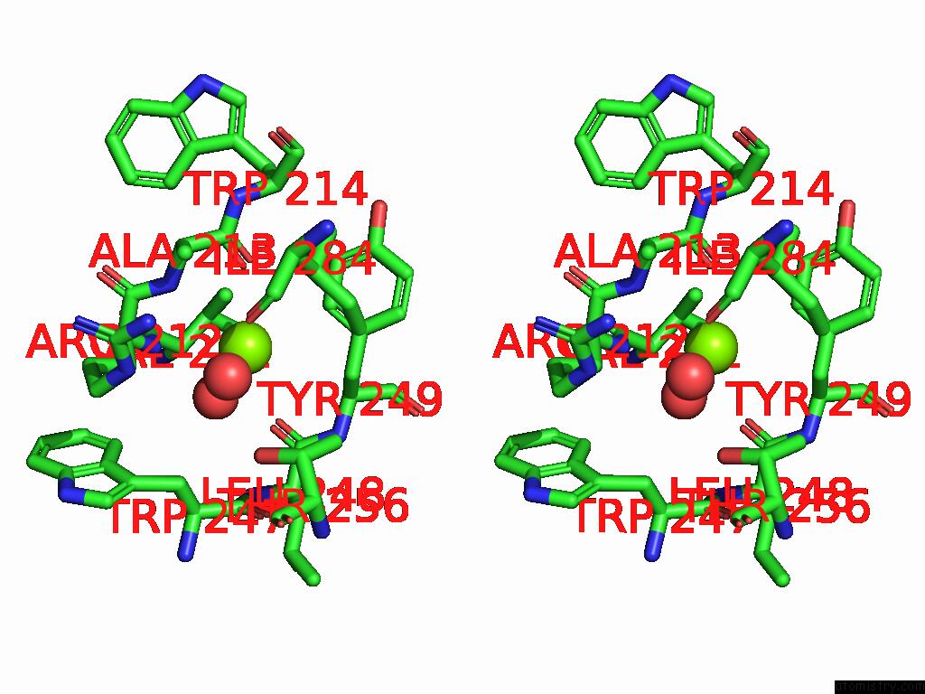
Magnesium binding site 2 out of 3 in 9bfa
Go back to
Magnesium binding site 2 out
of 3 in the Bcat Mutant
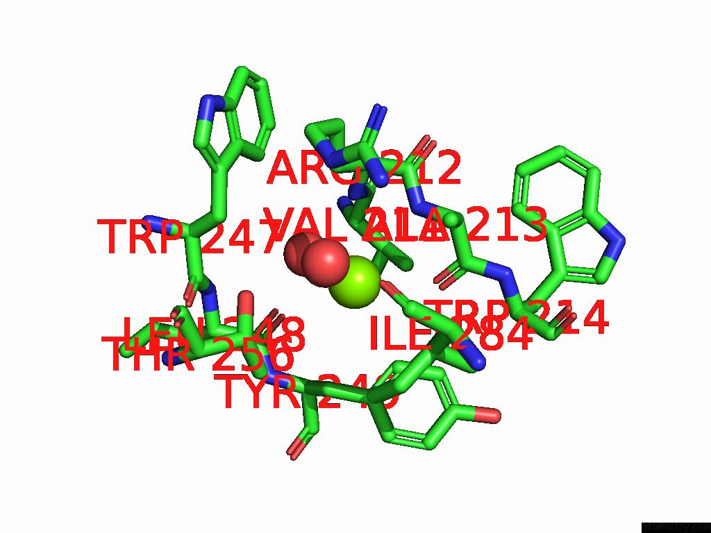
Mono view
Stereo pair view
Mono view
Stereo pair view
A full contact list of Magnesium with other atoms in the Mg binding
site number 2 of Bcat Mutant within 5.0Å range:
|
Magnesium binding site 3 out of 3 in 9bfa
Go back to
Magnesium binding site 3 out
of 3 in the Bcat Mutant
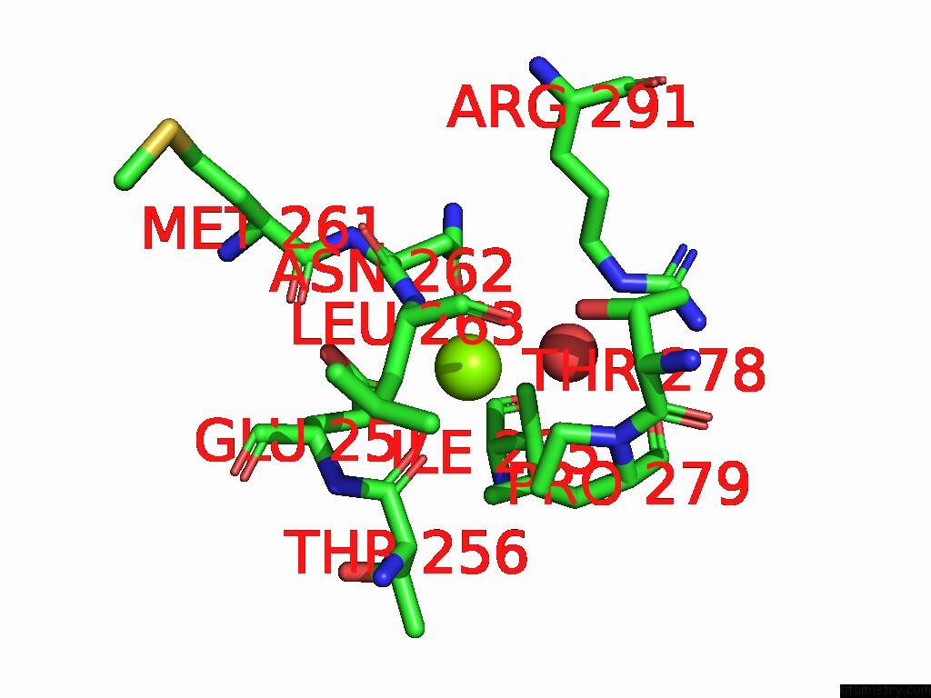
Mono view
Stereo pair view
Mono view
Stereo pair view
A full contact list of Magnesium with other atoms in the Mg binding
site number 3 of Bcat Mutant within 5.0Å range:
|
Reference:
E.S.Dare,
R.H.Newman,
M.E.Conway,
M.Dong.
Crystal Structures of the Phosphorylation Mimics of Human Cytosolic Branched Chain Aminotransferase. Arch.Biochem.Biophys. V. 770 10479 2025.
ISSN: ESSN 1096-0384
PubMed: 40414328
DOI: 10.1016/J.ABB.2025.110479
Page generated: Fri Aug 15 23:16:31 2025
ISSN: ESSN 1096-0384
PubMed: 40414328
DOI: 10.1016/J.ABB.2025.110479
Last articles
Na in 4OX2Na in 4OY6
Na in 4OWE
Na in 4OWH
Na in 4OQ9
Na in 4OWC
Na in 4OWB
Na in 4OVZ
Na in 4OUB
Na in 4OUA