Magnesium »
PDB 9chz-9dp4 »
9dfw »
Magnesium in PDB 9dfw: X-Ray Crystal Structure of An Engineered Viperin-Like Enzyme From T. Virens with Bound Ctp and Sam
Protein crystallography data
The structure of X-Ray Crystal Structure of An Engineered Viperin-Like Enzyme From T. Virens with Bound Ctp and Sam, PDB code: 9dfw
was solved by
J.C.Lachowicz,
J.B.Bonanno,
T.L.Grove,
with X-Ray Crystallography technique. A brief refinement statistics is given in the table below:
| Resolution Low / High (Å) | 19.86 / 1.68 |
| Space group | P 31 |
| Cell size a, b, c (Å), α, β, γ (°) | 85.131, 85.131, 110.471, 90, 90, 120 |
| R / Rfree (%) | 15.5 / 18.8 |
Other elements in 9dfw:
The structure of X-Ray Crystal Structure of An Engineered Viperin-Like Enzyme From T. Virens with Bound Ctp and Sam also contains other interesting chemical elements:
| Iron | (Fe) | 12 atoms |
Magnesium Binding Sites:
The binding sites of Magnesium atom in the X-Ray Crystal Structure of An Engineered Viperin-Like Enzyme From T. Virens with Bound Ctp and Sam
(pdb code 9dfw). This binding sites where shown within
5.0 Angstroms radius around Magnesium atom.
In total 3 binding sites of Magnesium where determined in the X-Ray Crystal Structure of An Engineered Viperin-Like Enzyme From T. Virens with Bound Ctp and Sam, PDB code: 9dfw:
Jump to Magnesium binding site number: 1; 2; 3;
In total 3 binding sites of Magnesium where determined in the X-Ray Crystal Structure of An Engineered Viperin-Like Enzyme From T. Virens with Bound Ctp and Sam, PDB code: 9dfw:
Jump to Magnesium binding site number: 1; 2; 3;
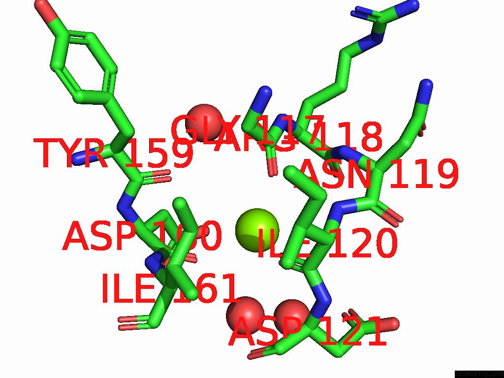
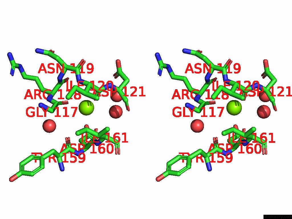
Magnesium binding site 1 out of 3 in 9dfw
Go back to
Magnesium binding site 1 out
of 3 in the X-Ray Crystal Structure of An Engineered Viperin-Like Enzyme From T. Virens with Bound Ctp and Sam
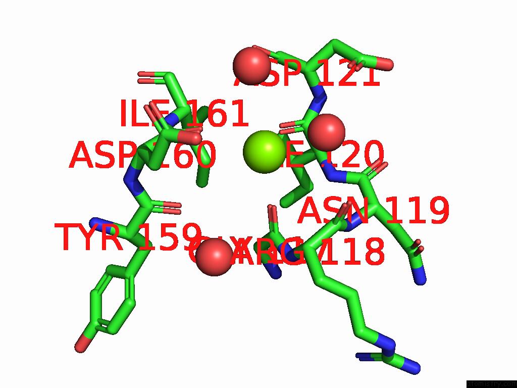
Mono view
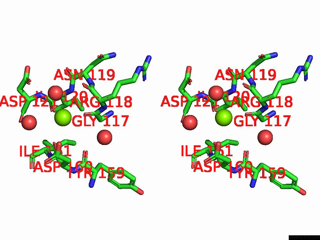
Stereo pair view
Mono view
Stereo pair view
A full contact list of Magnesium with other atoms in the Mg binding
site number 1 of X-Ray Crystal Structure of An Engineered Viperin-Like Enzyme From T. Virens with Bound Ctp and Sam within 5.0Å range:
|
Magnesium binding site 2 out of 3 in 9dfw
Go back to
Magnesium binding site 2 out
of 3 in the X-Ray Crystal Structure of An Engineered Viperin-Like Enzyme From T. Virens with Bound Ctp and Sam
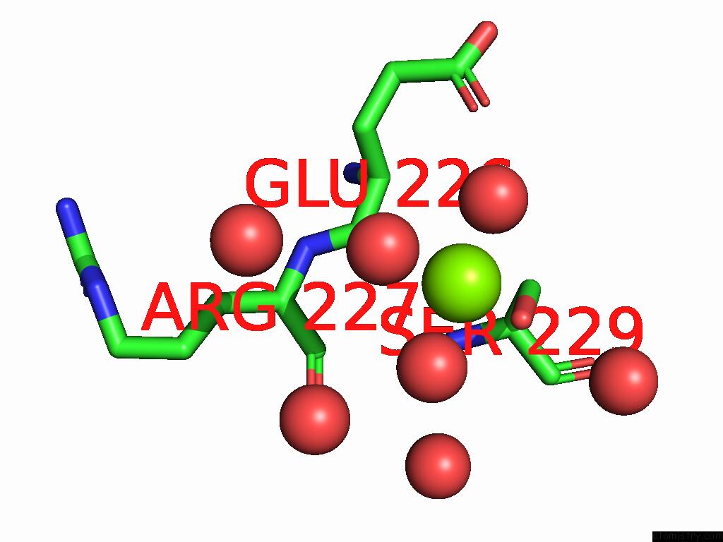
Mono view
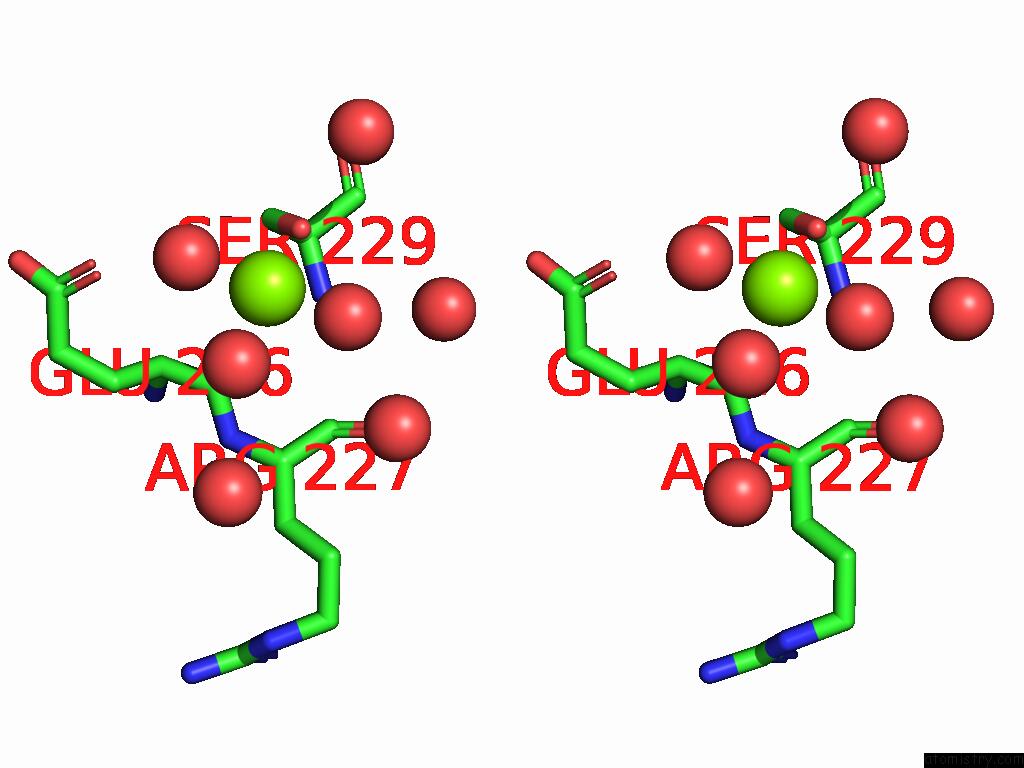
Stereo pair view
Mono view
Stereo pair view
A full contact list of Magnesium with other atoms in the Mg binding
site number 2 of X-Ray Crystal Structure of An Engineered Viperin-Like Enzyme From T. Virens with Bound Ctp and Sam within 5.0Å range:
|
Magnesium binding site 3 out of 3 in 9dfw
Go back to
Magnesium binding site 3 out
of 3 in the X-Ray Crystal Structure of An Engineered Viperin-Like Enzyme From T. Virens with Bound Ctp and Sam
Mono view
Stereo pair view
Mono view
Stereo pair view
A full contact list of Magnesium with other atoms in the Mg binding
site number 3 of X-Ray Crystal Structure of An Engineered Viperin-Like Enzyme From T. Virens with Bound Ctp and Sam within 5.0Å range:
|
Reference:
J.C.Lachowicz,
S.Grudman,
J.B.Bonanno,
A.Fiser,
T.L.Grove.
Structural Insights From Active Site Variants and B-8 Loop Interactions in Viperin-Like Enzymes To Be Published.
Page generated: Tue Feb 25 11:16:28 2025
Last articles
Fe in 2YXOFe in 2YRS
Fe in 2YXC
Fe in 2YNM
Fe in 2YVJ
Fe in 2YP1
Fe in 2YU2
Fe in 2YU1
Fe in 2YQB
Fe in 2YOO