Magnesium »
PDB 9dz6-9ei1 »
9e9b »
Magnesium in PDB 9e9b: Crystal Structure of L. Monocytogenes Mend with MG2+ and Thdp Bound
Enzymatic activity of Crystal Structure of L. Monocytogenes Mend with MG2+ and Thdp Bound
All present enzymatic activity of Crystal Structure of L. Monocytogenes Mend with MG2+ and Thdp Bound:
2.2.1.9;
2.2.1.9;
Protein crystallography data
The structure of Crystal Structure of L. Monocytogenes Mend with MG2+ and Thdp Bound, PDB code: 9e9b
was solved by
M.Klein,
F.M.Given,
N.A.T.Ho,
T.M.Allison,
J.M.Johnston,
with X-Ray Crystallography technique. A brief refinement statistics is given in the table below:
| Resolution Low / High (Å) | 47.57 / 2.61 |
| Space group | P 64 2 2 |
| Cell size a, b, c (Å), α, β, γ (°) | 176.025, 176.025, 100.144, 90, 90, 120 |
| R / Rfree (%) | 19.9 / 23.5 |
Magnesium Binding Sites:
The binding sites of Magnesium atom in the Crystal Structure of L. Monocytogenes Mend with MG2+ and Thdp Bound
(pdb code 9e9b). This binding sites where shown within
5.0 Angstroms radius around Magnesium atom.
In total only one binding site of Magnesium was determined in the Crystal Structure of L. Monocytogenes Mend with MG2+ and Thdp Bound, PDB code: 9e9b:
In total only one binding site of Magnesium was determined in the Crystal Structure of L. Monocytogenes Mend with MG2+ and Thdp Bound, PDB code: 9e9b:
Magnesium binding site 1 out of 1 in 9e9b
Go back to
Magnesium binding site 1 out
of 1 in the Crystal Structure of L. Monocytogenes Mend with MG2+ and Thdp Bound
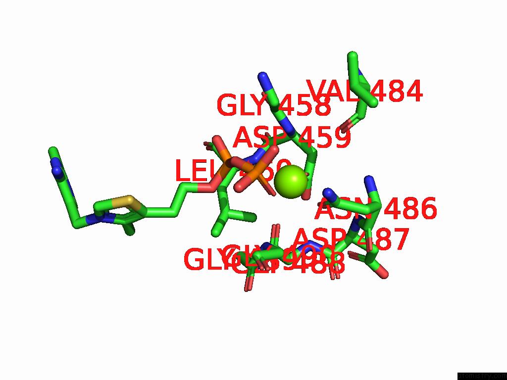
Mono view
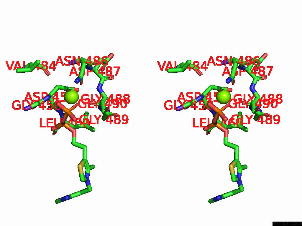
Stereo pair view
Mono view
Stereo pair view
A full contact list of Magnesium with other atoms in the Mg binding
site number 1 of Crystal Structure of L. Monocytogenes Mend with MG2+ and Thdp Bound within 5.0Å range:
|
Reference:
M.Bailey,
F.M.Given,
N.A.T.Ho,
F.G.Pearce,
T.M.Allison,
J.M.Johnston.
Structures of Listeria Monocytogenes Mend in Thdp-Bound and in-Crystallo Captured Intermediate I-Bound Forms. Acta Crystallogr.,Sect.F 2025.
ISSN: ESSN 2053-230X
PubMed: 40673487
DOI: 10.1107/S2053230X25006181
Page generated: Sat Aug 16 00:57:19 2025
ISSN: ESSN 2053-230X
PubMed: 40673487
DOI: 10.1107/S2053230X25006181
Last articles
Mn in 4UXAMn in 4W8Y
Mn in 4W9S
Mn in 4V15
Mn in 4V0U
Mn in 4V0W
Mn in 4V0X
Mn in 4UWQ
Mn in 4V0V
Mn in 4URR