Magnesium in PDB 9g6p: Xylose Isomerase Collected at 50C Using Time-Resolved Serial Synchrotron Crystallography with Glucose at 60 Seconds
Enzymatic activity of Xylose Isomerase Collected at 50C Using Time-Resolved Serial Synchrotron Crystallography with Glucose at 60 Seconds
All present enzymatic activity of Xylose Isomerase Collected at 50C Using Time-Resolved Serial Synchrotron Crystallography with Glucose at 60 Seconds:
5.3.1.5;
Protein crystallography data
The structure of Xylose Isomerase Collected at 50C Using Time-Resolved Serial Synchrotron Crystallography with Glucose at 60 Seconds, PDB code: 9g6p
was solved by
E.C.Schulz,
A.Prester,
D.V.Stetten,
G.Gore,
C.E.Hatton,
K.Bartels,
J.P.Leimkohl,
H.Schikora,
H.M.Ginn,
F.Tellkamp,
P.Mehrabi,
with X-Ray Crystallography technique. A brief refinement statistics is given in the table below:
Resolution Low / High (Å)
|
71.49 /
1.70
|
Space group
|
I 2 2 2
|
Cell size a, b, c (Å), α, β, γ (°)
|
94.2,
103.05,
99.25,
90,
90,
90
|
R / Rfree (%)
|
17.4 /
21.5
|
Magnesium Binding Sites:
The binding sites of Magnesium atom in the Xylose Isomerase Collected at 50C Using Time-Resolved Serial Synchrotron Crystallography with Glucose at 60 Seconds
(pdb code 9g6p). This binding sites where shown within
5.0 Angstroms radius around Magnesium atom.
In total 3 binding sites of Magnesium where determined in the
Xylose Isomerase Collected at 50C Using Time-Resolved Serial Synchrotron Crystallography with Glucose at 60 Seconds, PDB code: 9g6p:
Jump to Magnesium binding site number:
1;
2;
3;
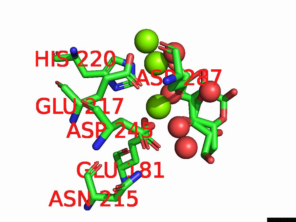
Magnesium binding site 1 out
of 3 in 9g6p
Go back to
Magnesium Binding Sites List in 9g6p
Magnesium binding site 1 out
of 3 in the Xylose Isomerase Collected at 50C Using Time-Resolved Serial Synchrotron Crystallography with Glucose at 60 Seconds
 Mono view
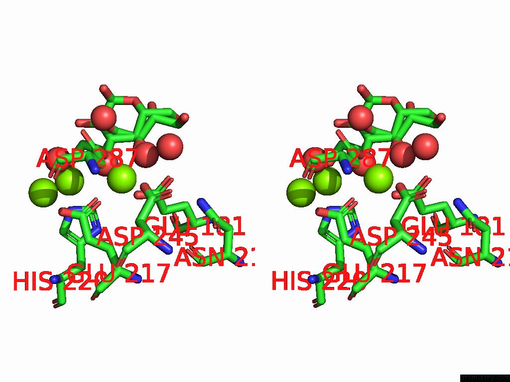
 Stereo pair view
|
A full contact list of Magnesium with other atoms in the Mg binding
site number 1 of Xylose Isomerase Collected at 50C Using Time-Resolved Serial Synchrotron Crystallography with Glucose at 60 Seconds within 5.0Å range:
probe
|
atom
|
residue
|
distance (Å)
|
B
|
Occ
|
A:Mg403
b:16.4
occ:0.96
|
OE1
|
A:GLU217
|
2.0
|
21.0
|
1.0
|
OE2
|
A:GLU181
|
2.0
|
22.5
|
1.0
|
OD2
|
A:ASP287
|
2.0
|
22.7
|
1.0
|
O
|
A:HOH515
|
2.0
|
22.4
|
0.3
|
O
|
A:HOH532
|
2.0
|
20.4
|
0.3
|
O4
|
A:GLO402
|
2.0
|
23.0
|
0.4
|
OD2
|
A:ASP245
|
2.1
|
20.6
|
1.0
|
O2
|
A:GLO402
|
2.3
|
20.3
|
0.4
|
O4
|
A:GLC401
|
2.4
|
22.2
|
0.2
|
O3
|
A:GLC401
|
2.6
|
20.9
|
0.2
|
CD
|
A:GLU181
|
3.0
|
22.5
|
1.0
|
C3
|
A:GLC401
|
3.0
|
20.8
|
0.2
|
CG
|
A:ASP287
|
3.1
|
19.5
|
1.0
|
C4
|
A:GLO402
|
3.2
|
23.1
|
0.4
|
CD
|
A:GLU217
|
3.2
|
15.1
|
1.0
|
C4
|
A:GLC401
|
3.2
|
22.9
|
0.2
|
CG
|
A:ASP245
|
3.2
|
17.6
|
1.0
|
OE1
|
A:GLU181
|
3.3
|
22.0
|
1.0
|
C2
|
A:GLO402
|
3.4
|
22.3
|
0.4
|
MG
|
A:MG404
|
3.5
|
32.2
|
1.0
|
O3
|
A:GLO402
|
3.5
|
22.2
|
0.4
|
C3
|
A:GLO402
|
3.6
|
19.3
|
0.4
|
CB
|
A:ASP287
|
3.6
|
16.1
|
1.0
|
O
|
A:HOH518
|
3.7
|
37.1
|
1.0
|
O
|
A:HOH549
|
3.8
|
24.8
|
0.3
|
CB
|
A:ASP245
|
3.8
|
14.6
|
1.0
|
O
|
A:HOH512
|
4.0
|
19.9
|
1.0
|
OE2
|
A:GLU217
|
4.0
|
19.1
|
1.0
|
CE1
|
A:HIS220
|
4.1
|
16.4
|
1.0
|
CG
|
A:GLU217
|
4.2
|
14.8
|
1.0
|
OD1
|
A:ASP287
|
4.2
|
18.2
|
1.0
|
CB
|
A:GLU217
|
4.2
|
15.1
|
1.0
|
OD1
|
A:ASP245
|
4.3
|
18.3
|
1.0
|
CG
|
A:GLU181
|
4.3
|
15.0
|
1.0
|
C5
|
A:GLO402
|
4.4
|
19.1
|
0.4
|
C5
|
A:GLC401
|
4.5
|
22.2
|
0.2
|
C2
|
A:GLC401
|
4.6
|
22.2
|
0.2
|
NE2
|
A:HIS220
|
4.6
|
19.7
|
1.0
|
C1
|
A:GLO402
|
4.7
|
18.0
|
0.4
|
O1
|
A:GLO402
|
4.8
|
17.8
|
0.4
|
C6
|
A:GLO402
|
4.9
|
22.1
|
0.4
|
ND1
|
A:HIS220
|
4.9
|
17.7
|
1.0
|
ND2
|
A:ASN215
|
4.9
|
16.1
|
1.0
|
MG
|
A:MG405
|
5.0
|
20.5
|
1.0
|
|
Magnesium binding site 2 out
of 3 in 9g6p
Go back to
Magnesium Binding Sites List in 9g6p
Magnesium binding site 2 out
of 3 in the Xylose Isomerase Collected at 50C Using Time-Resolved Serial Synchrotron Crystallography with Glucose at 60 Seconds
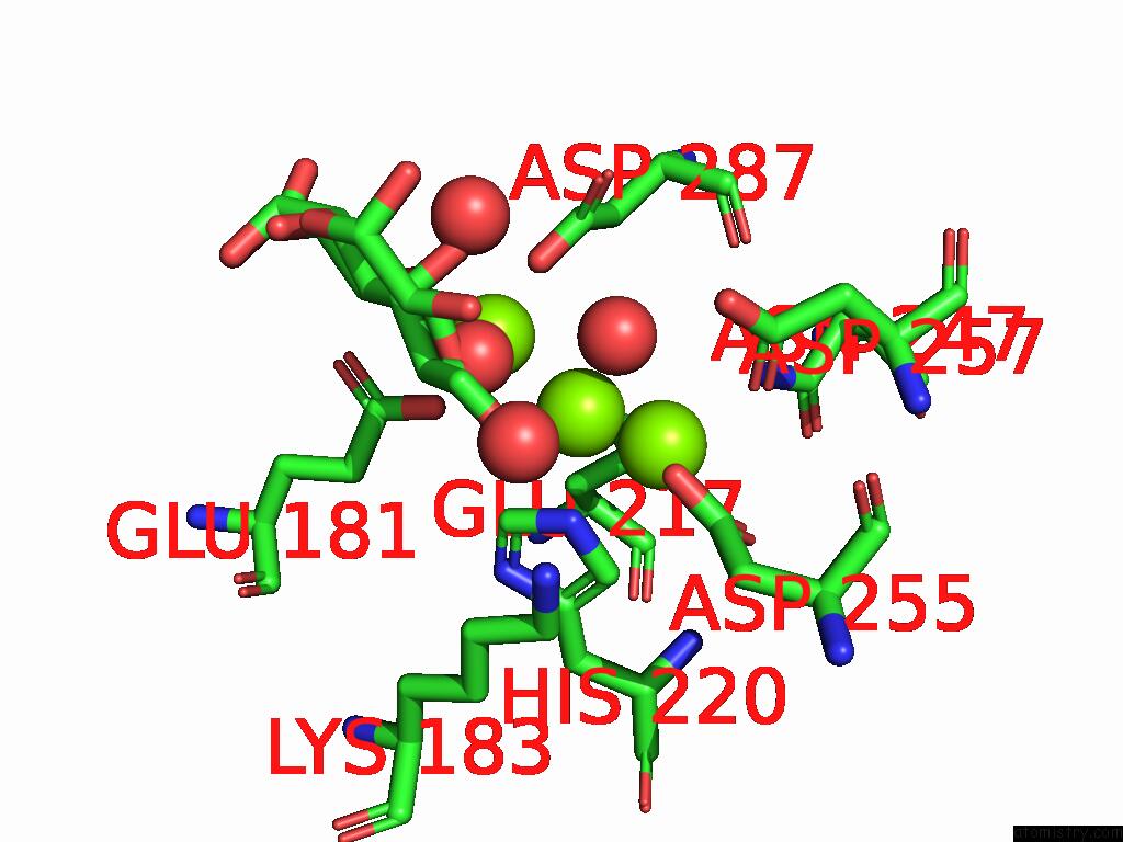
 Mono view
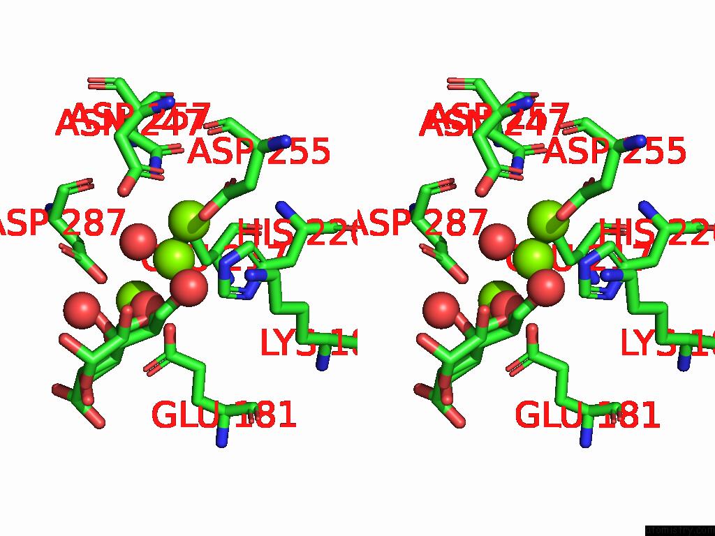
 Stereo pair view
|
A full contact list of Magnesium with other atoms in the Mg binding
site number 2 of Xylose Isomerase Collected at 50C Using Time-Resolved Serial Synchrotron Crystallography with Glucose at 60 Seconds within 5.0Å range:
probe
|
atom
|
residue
|
distance (Å)
|
B
|
Occ
|
A:Mg404
b:32.2
occ:0.96
|
MG
|
A:MG405
|
1.7
|
20.5
|
1.0
|
O
|
A:HOH512
|
1.8
|
19.9
|
1.0
|
O2
|
A:GLO402
|
2.2
|
20.3
|
0.4
|
NE2
|
A:HIS220
|
2.2
|
19.7
|
1.0
|
O1
|
A:GLO402
|
2.3
|
17.8
|
0.4
|
OE2
|
A:GLU217
|
2.3
|
19.1
|
1.0
|
O
|
A:HOH532
|
2.4
|
20.4
|
0.3
|
O3
|
A:GLC401
|
2.6
|
20.9
|
0.2
|
OE1
|
A:GLU217
|
2.7
|
21.0
|
1.0
|
CE1
|
A:HIS220
|
2.8
|
16.4
|
1.0
|
CD
|
A:GLU217
|
2.8
|
15.1
|
1.0
|
O
|
A:HOH637
|
2.8
|
21.0
|
0.3
|
OD2
|
A:ASP255
|
3.0
|
26.8
|
1.0
|
C1
|
A:GLO402
|
3.1
|
18.0
|
0.4
|
C2
|
A:GLO402
|
3.1
|
22.3
|
0.4
|
CD2
|
A:HIS220
|
3.4
|
16.0
|
1.0
|
MG
|
A:MG403
|
3.5
|
16.4
|
1.0
|
OD1
|
A:ASP257
|
3.6
|
20.4
|
1.0
|
OD2
|
A:ASP287
|
3.6
|
22.7
|
1.0
|
C3
|
A:GLC401
|
3.6
|
20.8
|
0.2
|
O2
|
A:GLC401
|
3.8
|
19.0
|
0.2
|
OD2
|
A:ASP257
|
3.9
|
20.9
|
1.0
|
CG
|
A:ASP255
|
3.9
|
23.9
|
1.0
|
ND1
|
A:HIS220
|
4.0
|
17.7
|
1.0
|
O3
|
A:GLO402
|
4.1
|
22.2
|
0.4
|
OD1
|
A:ASP255
|
4.1
|
23.5
|
1.0
|
CG
|
A:ASP257
|
4.2
|
19.5
|
1.0
|
C3
|
A:GLO402
|
4.2
|
19.3
|
0.4
|
C2
|
A:GLC401
|
4.3
|
22.2
|
0.2
|
OE2
|
A:GLU181
|
4.3
|
22.5
|
1.0
|
CG
|
A:GLU217
|
4.3
|
14.8
|
1.0
|
CG
|
A:HIS220
|
4.3
|
12.7
|
1.0
|
CG
|
A:ASP287
|
4.4
|
19.5
|
1.0
|
O
|
A:HOH549
|
4.4
|
24.8
|
0.3
|
O4
|
A:GLO402
|
4.6
|
23.0
|
0.4
|
ND2
|
A:ASN247
|
4.8
|
16.4
|
1.0
|
OD1
|
A:ASP287
|
4.8
|
18.2
|
1.0
|
C4
|
A:GLC401
|
4.9
|
22.9
|
0.2
|
CE
|
A:LYS183
|
4.9
|
18.8
|
1.0
|
CB
|
A:GLU217
|
4.9
|
15.1
|
1.0
|
NZ
|
A:LYS183
|
5.0
|
21.5
|
1.0
|
|
Magnesium binding site 3 out
of 3 in 9g6p
Go back to
Magnesium Binding Sites List in 9g6p
Magnesium binding site 3 out
of 3 in the Xylose Isomerase Collected at 50C Using Time-Resolved Serial Synchrotron Crystallography with Glucose at 60 Seconds
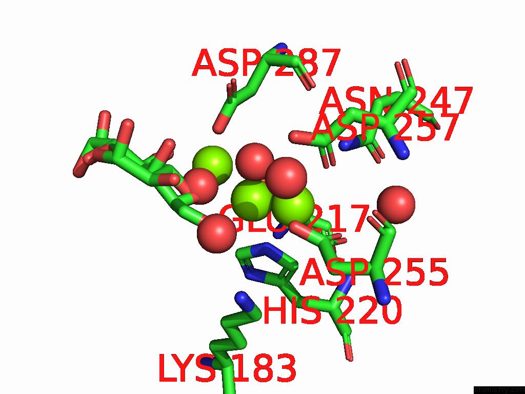
 Mono view
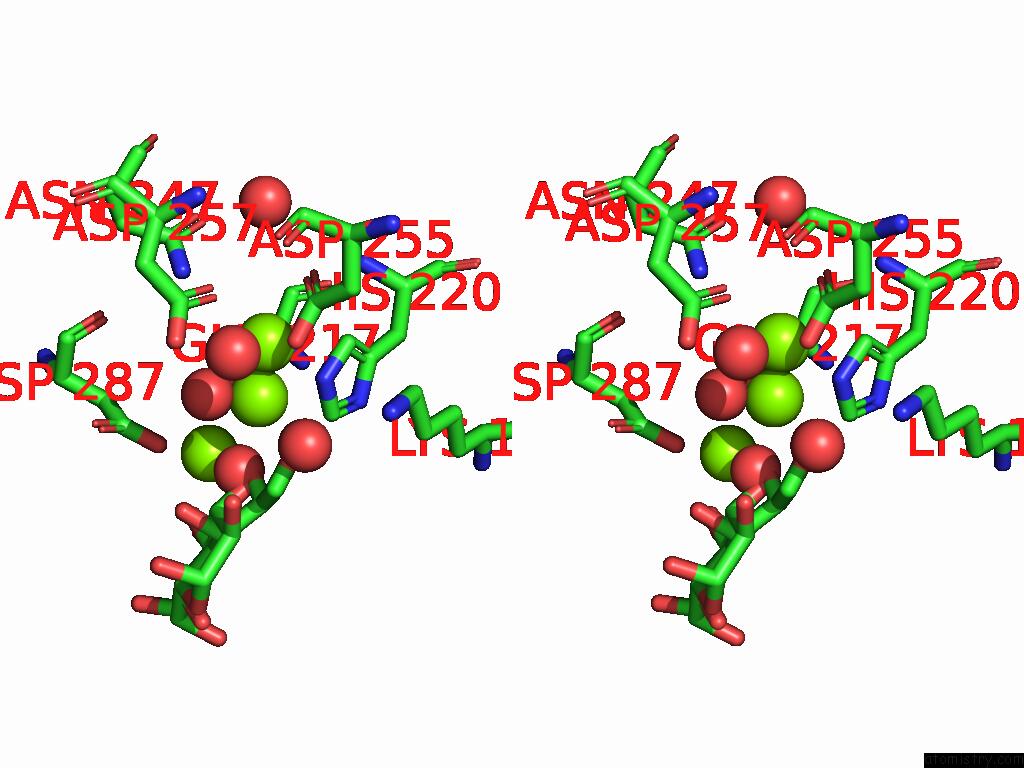
 Stereo pair view
|
A full contact list of Magnesium with other atoms in the Mg binding
site number 3 of Xylose Isomerase Collected at 50C Using Time-Resolved Serial Synchrotron Crystallography with Glucose at 60 Seconds within 5.0Å range:
probe
|
atom
|
residue
|
distance (Å)
|
B
|
Occ
|
A:Mg405
b:20.5
occ:0.96
|
MG
|
A:MG404
|
1.7
|
32.2
|
1.0
|
OD2
|
A:ASP255
|
1.9
|
26.8
|
1.0
|
OE2
|
A:GLU217
|
2.0
|
19.1
|
1.0
|
OD1
|
A:ASP257
|
2.3
|
20.4
|
1.0
|
O
|
A:HOH512
|
2.3
|
19.9
|
1.0
|
OD1
|
A:ASP255
|
2.4
|
23.5
|
1.0
|
CG
|
A:ASP255
|
2.5
|
23.9
|
1.0
|
NE2
|
A:HIS220
|
2.5
|
19.7
|
1.0
|
CD2
|
A:HIS220
|
3.1
|
16.0
|
1.0
|
CD
|
A:GLU217
|
3.1
|
15.1
|
1.0
|
CG
|
A:ASP257
|
3.1
|
19.5
|
1.0
|
O1
|
A:GLO402
|
3.3
|
17.8
|
0.4
|
OD2
|
A:ASP257
|
3.3
|
20.9
|
1.0
|
O
|
A:HOH637
|
3.5
|
21.0
|
0.3
|
CE1
|
A:HIS220
|
3.6
|
16.4
|
1.0
|
OE1
|
A:GLU217
|
3.6
|
21.0
|
1.0
|
O2
|
A:GLO402
|
3.9
|
20.3
|
0.4
|
CB
|
A:ASP255
|
4.0
|
20.7
|
1.0
|
ND2
|
A:ASN247
|
4.0
|
16.4
|
1.0
|
O
|
A:HOH589
|
4.2
|
19.4
|
1.0
|
O
|
A:HOH532
|
4.2
|
20.4
|
0.3
|
CG
|
A:HIS220
|
4.3
|
12.7
|
1.0
|
O3
|
A:GLC401
|
4.4
|
20.9
|
0.2
|
CG
|
A:GLU217
|
4.4
|
14.8
|
1.0
|
C1
|
A:GLO402
|
4.4
|
18.0
|
0.4
|
ND1
|
A:HIS220
|
4.6
|
17.7
|
1.0
|
O
|
A:HOH644
|
4.6
|
33.8
|
1.0
|
CB
|
A:ASP257
|
4.6
|
14.9
|
1.0
|
CE
|
A:LYS183
|
4.7
|
18.8
|
1.0
|
C2
|
A:GLO402
|
4.8
|
22.3
|
0.4
|
NZ
|
A:LYS183
|
4.8
|
21.5
|
1.0
|
OD2
|
A:ASP287
|
4.9
|
22.7
|
1.0
|
MG
|
A:MG403
|
5.0
|
16.4
|
1.0
|
CA
|
A:ASP255
|
5.0
|
14.3
|
1.0
|
CA
|
A:ASP257
|
5.0
|
15.8
|
1.0
|
|
Reference:
E.C.Schulz,
A.Prester,
D.Von Stetten,
G.Gore,
C.E.Hatton,
K.Bartels,
J.P.Leimkohl,
H.Schikora,
H.M.Ginn,
F.Tellkamp,
P.Mehrabi.
Probing the Modulation of Enzyme Kinetics By Multi-Temperature, Time-Resolved Serial Crystallography. Nat Commun V. 16 6553 2025.
ISSN: ESSN 2041-1723
PubMed: 40670369
DOI: 10.1038/S41467-025-61631-2
Page generated: Sat Aug 16 02:55:36 2025
|