Magnesium »
PDB 9gch-9h4e »
9gre »
Magnesium in PDB 9gre: Cryo-Electron Microscopy Structure of Glucose/Xylose Isomerase From Streptomyces Rubiginosus with Magnesium Ions in the Active Site
Enzymatic activity of Cryo-Electron Microscopy Structure of Glucose/Xylose Isomerase From Streptomyces Rubiginosus with Magnesium Ions in the Active Site
All present enzymatic activity of Cryo-Electron Microscopy Structure of Glucose/Xylose Isomerase From Streptomyces Rubiginosus with Magnesium Ions in the Active Site:
5.3.1.5;
5.3.1.5;
Magnesium Binding Sites:
The binding sites of Magnesium atom in the Cryo-Electron Microscopy Structure of Glucose/Xylose Isomerase From Streptomyces Rubiginosus with Magnesium Ions in the Active Site
(pdb code 9gre). This binding sites where shown within
5.0 Angstroms radius around Magnesium atom.
In total 8 binding sites of Magnesium where determined in the Cryo-Electron Microscopy Structure of Glucose/Xylose Isomerase From Streptomyces Rubiginosus with Magnesium Ions in the Active Site, PDB code: 9gre:
Jump to Magnesium binding site number: 1; 2; 3; 4; 5; 6; 7; 8;
In total 8 binding sites of Magnesium where determined in the Cryo-Electron Microscopy Structure of Glucose/Xylose Isomerase From Streptomyces Rubiginosus with Magnesium Ions in the Active Site, PDB code: 9gre:
Jump to Magnesium binding site number: 1; 2; 3; 4; 5; 6; 7; 8;
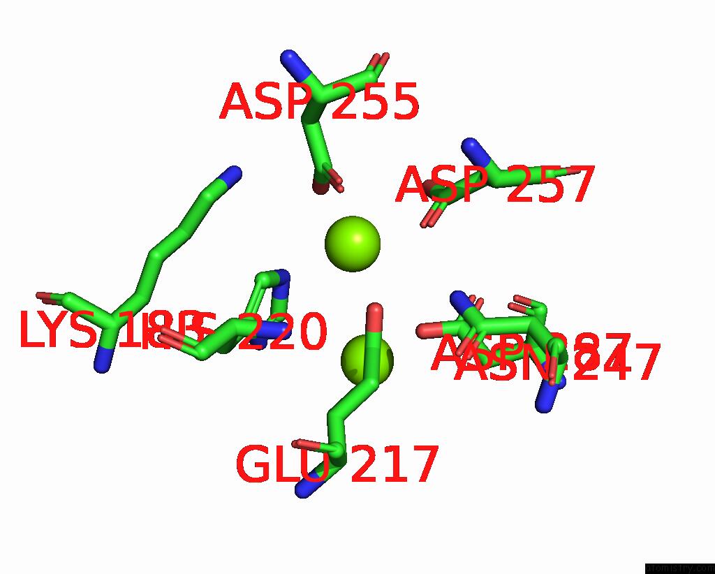
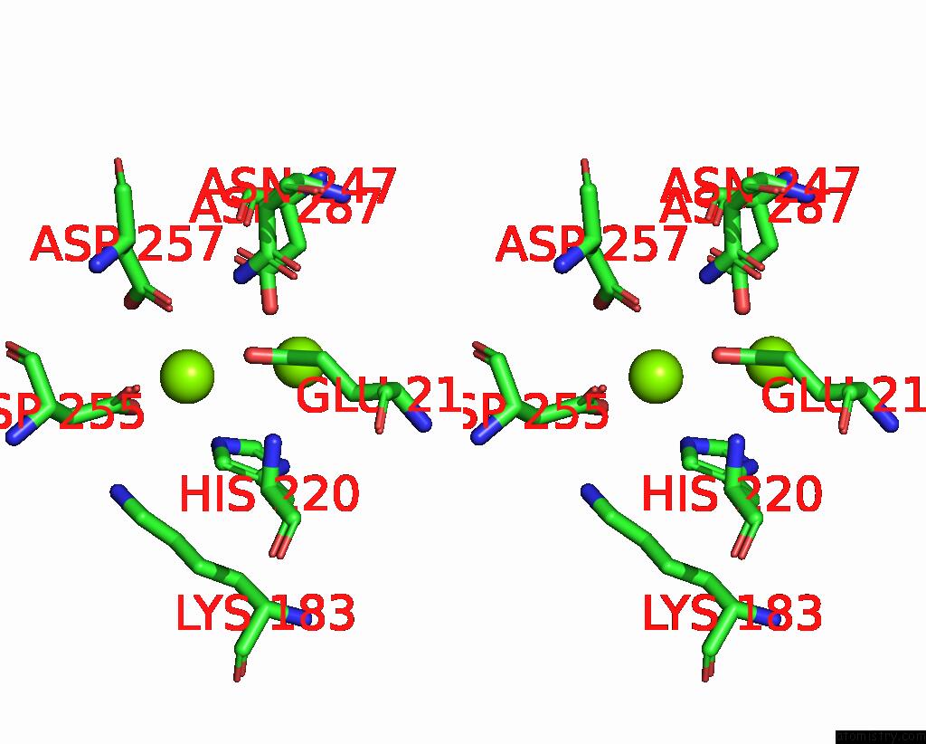
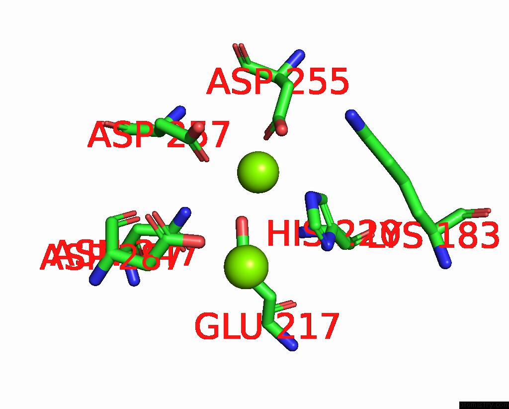
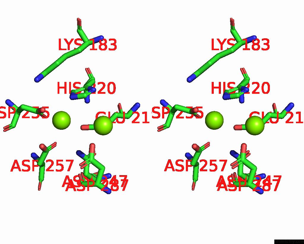
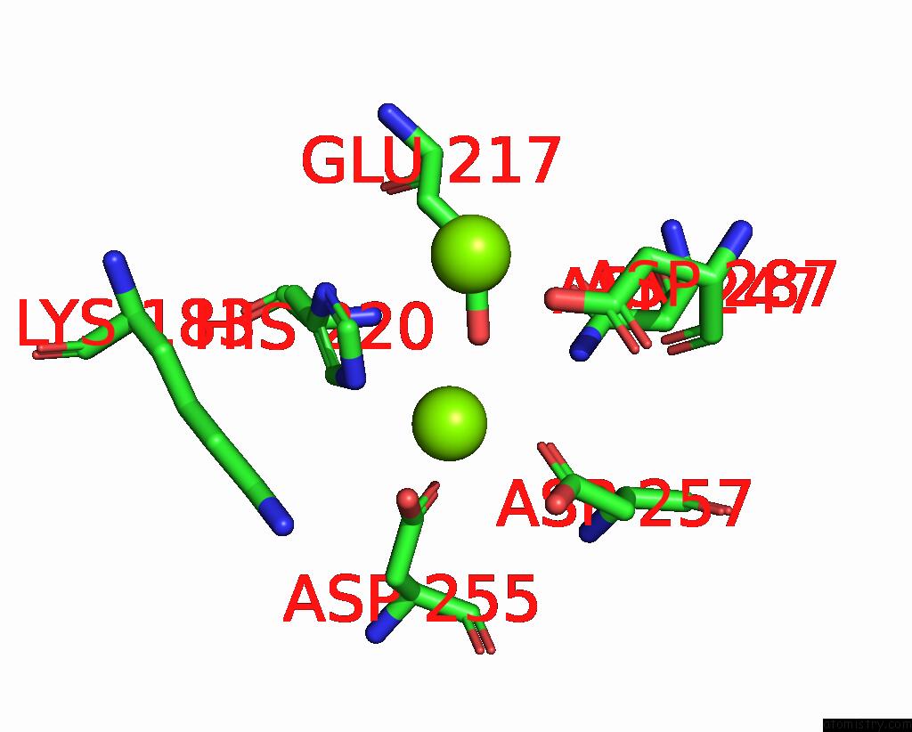
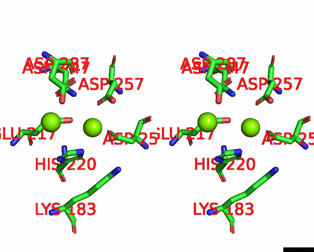
Magnesium binding site 1 out of 8 in 9gre
Go back to
Magnesium binding site 1 out
of 8 in the Cryo-Electron Microscopy Structure of Glucose/Xylose Isomerase From Streptomyces Rubiginosus with Magnesium Ions in the Active Site
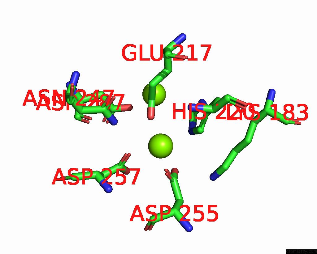
Mono view
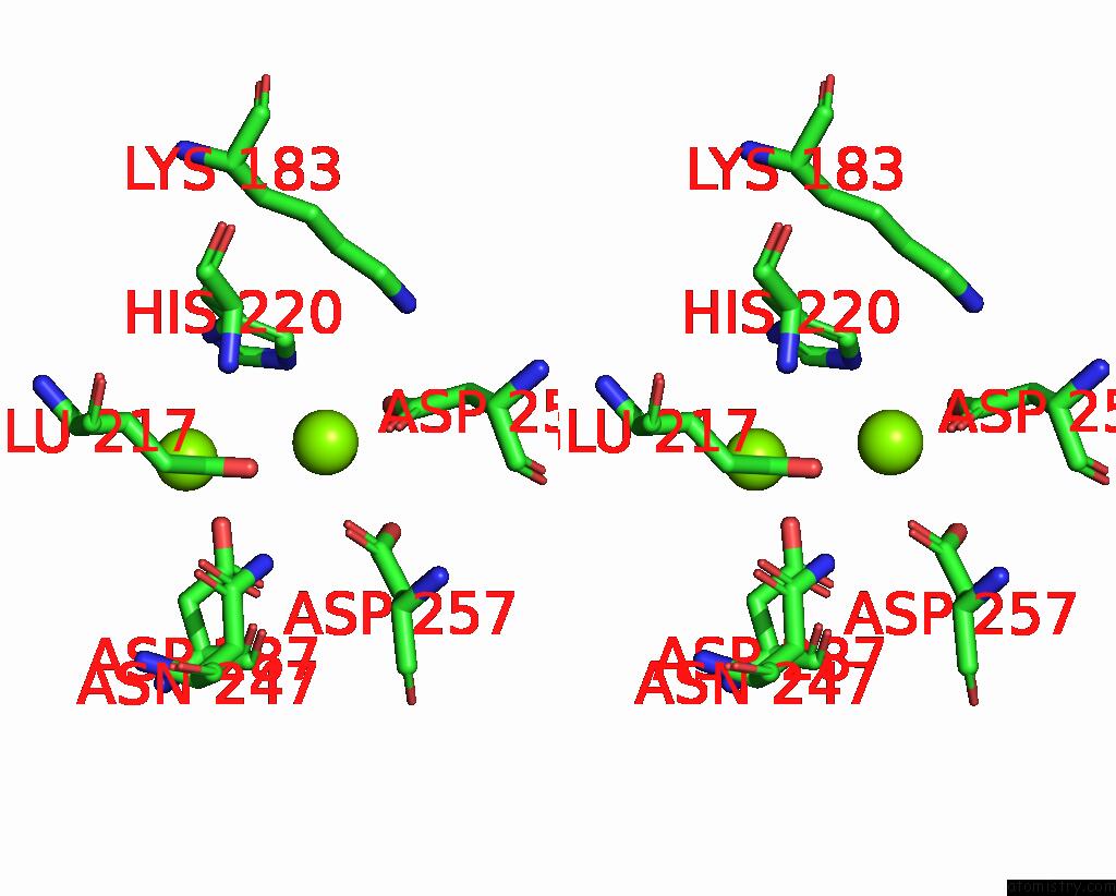
Stereo pair view
Mono view
Stereo pair view
A full contact list of Magnesium with other atoms in the Mg binding
site number 1 of Cryo-Electron Microscopy Structure of Glucose/Xylose Isomerase From Streptomyces Rubiginosus with Magnesium Ions in the Active Site within 5.0Å range:
|
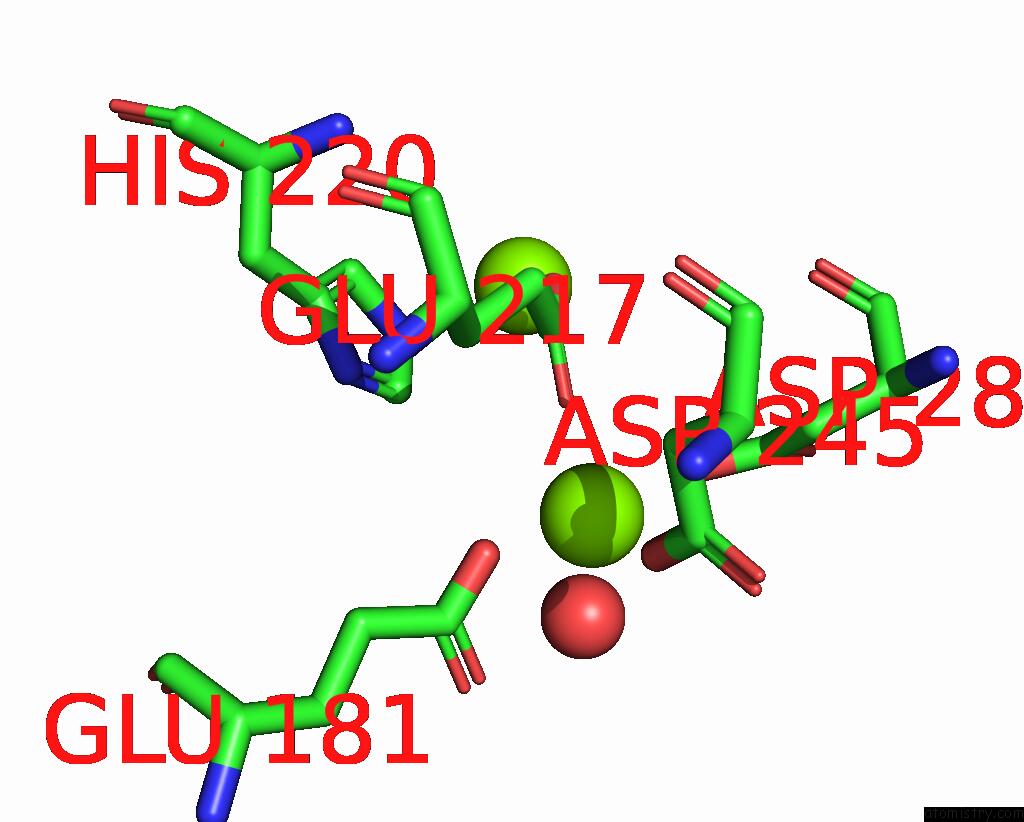
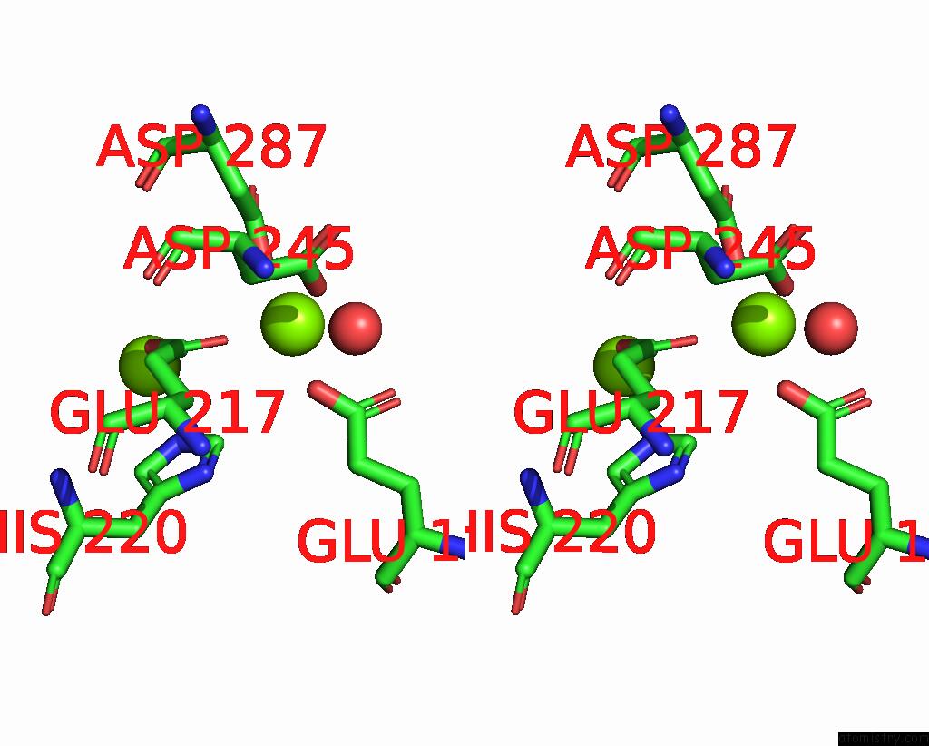
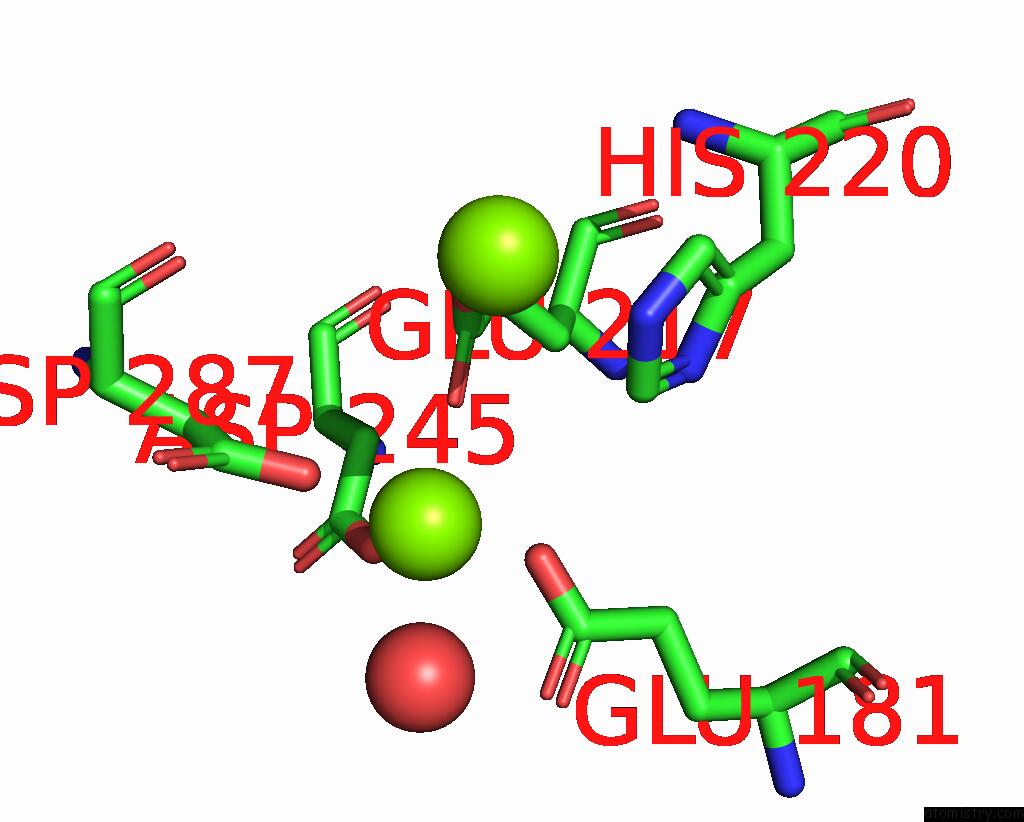
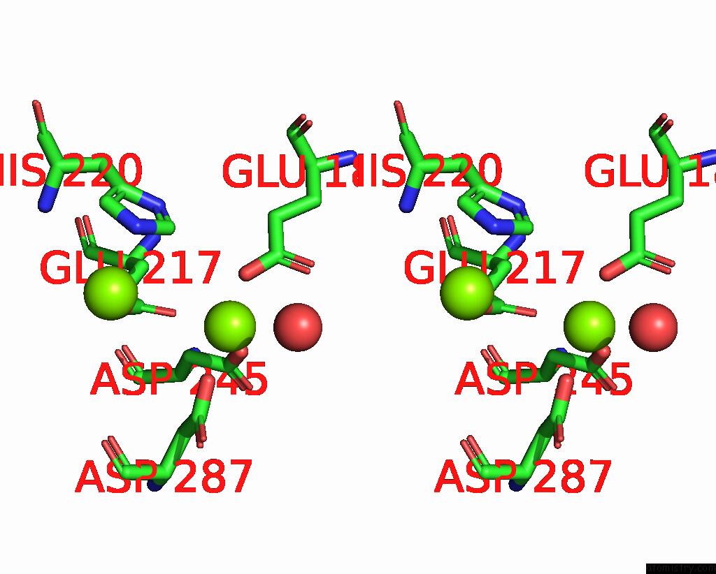
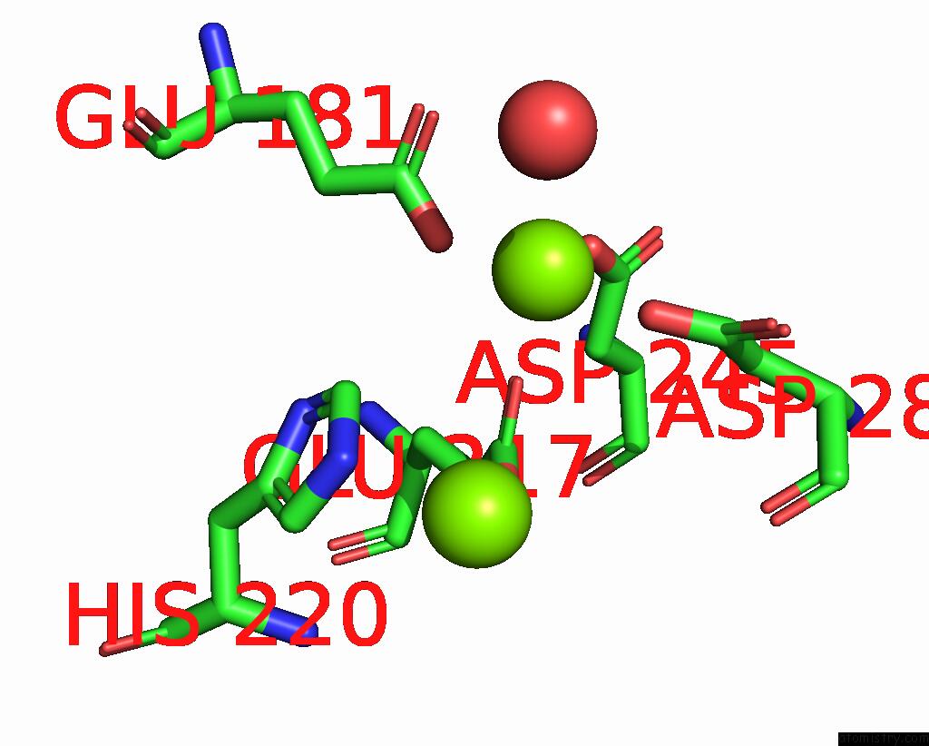
Magnesium binding site 2 out of 8 in 9gre
Go back to
Magnesium binding site 2 out
of 8 in the Cryo-Electron Microscopy Structure of Glucose/Xylose Isomerase From Streptomyces Rubiginosus with Magnesium Ions in the Active Site
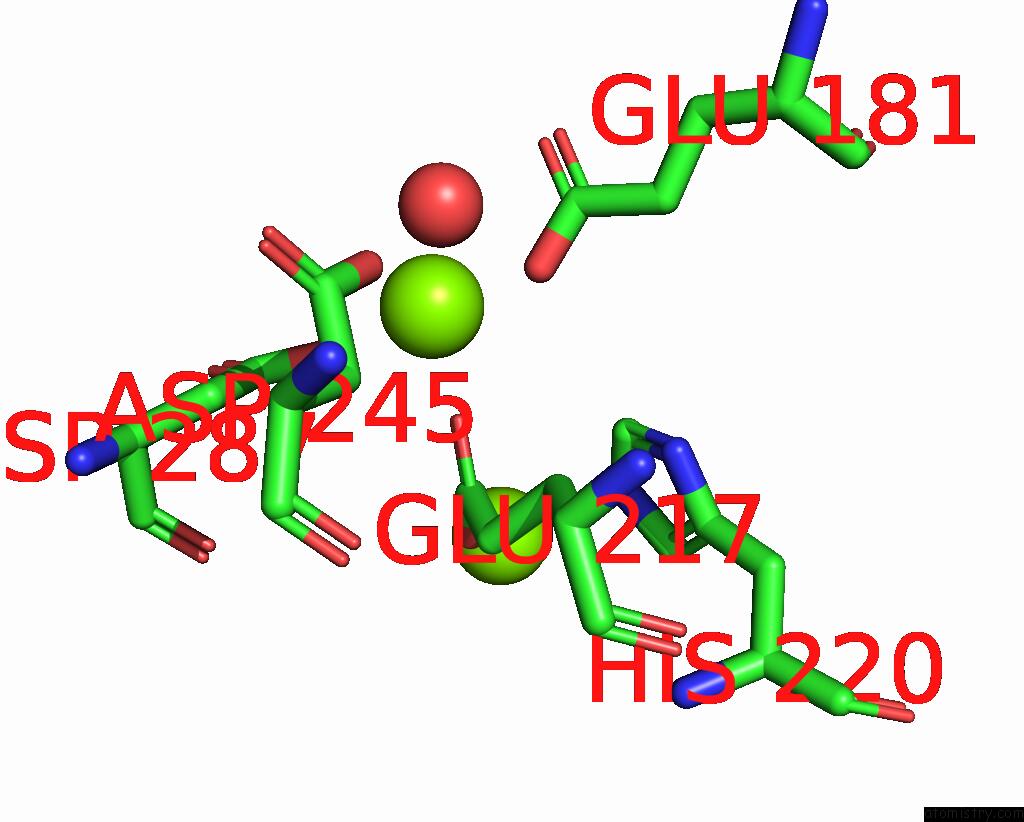
Mono view
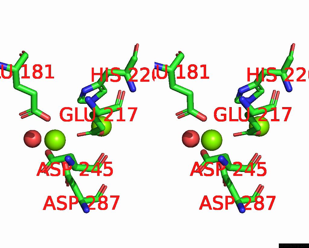
Stereo pair view
Mono view
Stereo pair view
A full contact list of Magnesium with other atoms in the Mg binding
site number 2 of Cryo-Electron Microscopy Structure of Glucose/Xylose Isomerase From Streptomyces Rubiginosus with Magnesium Ions in the Active Site within 5.0Å range:
|
Magnesium binding site 3 out of 8 in 9gre
Go back to
Magnesium binding site 3 out
of 8 in the Cryo-Electron Microscopy Structure of Glucose/Xylose Isomerase From Streptomyces Rubiginosus with Magnesium Ions in the Active Site
Mono view
Stereo pair view
Mono view
Stereo pair view
A full contact list of Magnesium with other atoms in the Mg binding
site number 3 of Cryo-Electron Microscopy Structure of Glucose/Xylose Isomerase From Streptomyces Rubiginosus with Magnesium Ions in the Active Site within 5.0Å range:
|
Magnesium binding site 4 out of 8 in 9gre
Go back to
Magnesium binding site 4 out
of 8 in the Cryo-Electron Microscopy Structure of Glucose/Xylose Isomerase From Streptomyces Rubiginosus with Magnesium Ions in the Active Site
Mono view
Stereo pair view
Mono view
Stereo pair view
A full contact list of Magnesium with other atoms in the Mg binding
site number 4 of Cryo-Electron Microscopy Structure of Glucose/Xylose Isomerase From Streptomyces Rubiginosus with Magnesium Ions in the Active Site within 5.0Å range:
|
Magnesium binding site 5 out of 8 in 9gre
Go back to
Magnesium binding site 5 out
of 8 in the Cryo-Electron Microscopy Structure of Glucose/Xylose Isomerase From Streptomyces Rubiginosus with Magnesium Ions in the Active Site
Mono view
Stereo pair view
Mono view
Stereo pair view
A full contact list of Magnesium with other atoms in the Mg binding
site number 5 of Cryo-Electron Microscopy Structure of Glucose/Xylose Isomerase From Streptomyces Rubiginosus with Magnesium Ions in the Active Site within 5.0Å range:
|
Magnesium binding site 6 out of 8 in 9gre
Go back to
Magnesium binding site 6 out
of 8 in the Cryo-Electron Microscopy Structure of Glucose/Xylose Isomerase From Streptomyces Rubiginosus with Magnesium Ions in the Active Site
Mono view
Stereo pair view
Mono view
Stereo pair view
A full contact list of Magnesium with other atoms in the Mg binding
site number 6 of Cryo-Electron Microscopy Structure of Glucose/Xylose Isomerase From Streptomyces Rubiginosus with Magnesium Ions in the Active Site within 5.0Å range:
|
Magnesium binding site 7 out of 8 in 9gre
Go back to
Magnesium binding site 7 out
of 8 in the Cryo-Electron Microscopy Structure of Glucose/Xylose Isomerase From Streptomyces Rubiginosus with Magnesium Ions in the Active Site
Mono view
Stereo pair view
Mono view
Stereo pair view
A full contact list of Magnesium with other atoms in the Mg binding
site number 7 of Cryo-Electron Microscopy Structure of Glucose/Xylose Isomerase From Streptomyces Rubiginosus with Magnesium Ions in the Active Site within 5.0Å range:
|
Magnesium binding site 8 out of 8 in 9gre
Go back to
Magnesium binding site 8 out
of 8 in the Cryo-Electron Microscopy Structure of Glucose/Xylose Isomerase From Streptomyces Rubiginosus with Magnesium Ions in the Active Site
Mono view

Stereo pair view
Mono view
Stereo pair view
A full contact list of Magnesium with other atoms in the Mg binding
site number 8 of Cryo-Electron Microscopy Structure of Glucose/Xylose Isomerase From Streptomyces Rubiginosus with Magnesium Ions in the Active Site within 5.0Å range:
|
Reference:
J.Slawek,
A.Klonecka,
M.Rawski,
M.Kozak.
Cryo-Electron Microscopy Structure of Glucose/Xylose Isomerase From Streptomyces Rubiginosus with Magnesium Ions in the Active Site To Be Published.
Page generated: Sat Aug 16 03:05:52 2025
Last articles
Mg in 9LK4Mg in 9LK5
Mg in 9KQQ
Mg in 9MF6
Mg in 9MED
Mg in 9MEC
Mg in 9MDY
Mg in 9MDW
Mg in 9M84
Mg in 9M7M