Magnesium »
PDB 9nf0-9qof »
9o3q »
Magnesium in PDB 9o3q: Apo-Structure of A Chondroitinase
Protein crystallography data
The structure of Apo-Structure of A Chondroitinase, PDB code: 9o3q
was solved by
B.Alvarez,
A.B.Boraston,
with X-Ray Crystallography technique. A brief refinement statistics is given in the table below:
| Resolution Low / High (Å) | 29.52 / 1.75 |
| Space group | P 21 21 21 |
| Cell size a, b, c (Å), α, β, γ (°) | 69.284, 84.043, 112.87, 90, 90, 90 |
| R / Rfree (%) | 18.5 / 21.8 |
Magnesium Binding Sites:
The binding sites of Magnesium atom in the Apo-Structure of A Chondroitinase
(pdb code 9o3q). This binding sites where shown within
5.0 Angstroms radius around Magnesium atom.
In total 2 binding sites of Magnesium where determined in the Apo-Structure of A Chondroitinase, PDB code: 9o3q:
Jump to Magnesium binding site number: 1; 2;
In total 2 binding sites of Magnesium where determined in the Apo-Structure of A Chondroitinase, PDB code: 9o3q:
Jump to Magnesium binding site number: 1; 2;
Magnesium binding site 1 out of 2 in 9o3q
Go back to
Magnesium binding site 1 out
of 2 in the Apo-Structure of A Chondroitinase
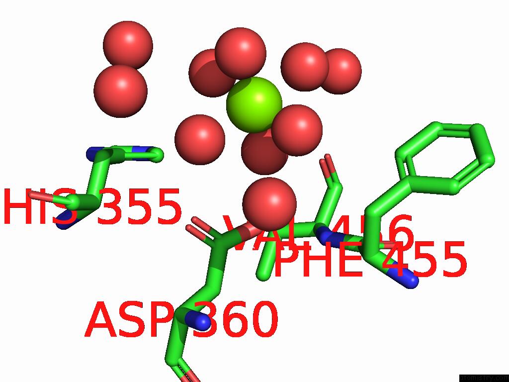
Mono view
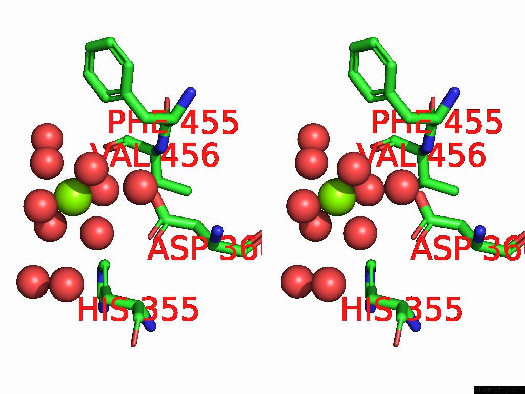
Stereo pair view
Mono view
Stereo pair view
A full contact list of Magnesium with other atoms in the Mg binding
site number 1 of Apo-Structure of A Chondroitinase within 5.0Å range:
|
Magnesium binding site 2 out of 2 in 9o3q
Go back to
Magnesium binding site 2 out
of 2 in the Apo-Structure of A Chondroitinase
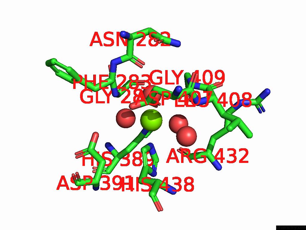
Mono view
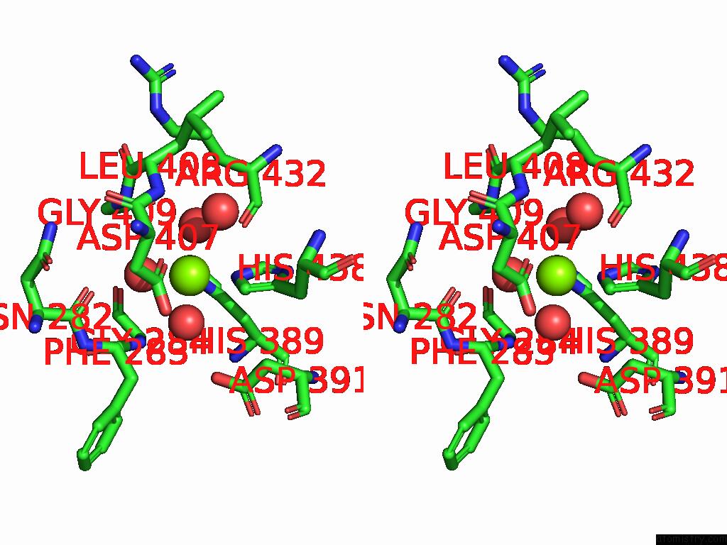
Stereo pair view
Mono view
Stereo pair view
A full contact list of Magnesium with other atoms in the Mg binding
site number 2 of Apo-Structure of A Chondroitinase within 5.0Å range:
|
Reference:
B.Alvarez,
O.F.Canil,
K.L.Low,
W.D.Abbott,
A.B.Boraston.
Analysis of Chondroitin Degradation By Components of A Bacteroides Caccae Polysaccharide Utilization Locus To Be Published.
Page generated: Sat Aug 16 06:57:43 2025
Last articles
Mn in 2AECMn in 2AE7
Mn in 2ADQ
Mn in 2A8S
Mn in 2ADP
Mn in 2A9P
Mn in 2A9O
Mn in 2A9I
Mn in 2A8W
Mn in 2A8U