Magnesium »
PDB 9nf0-9qof »
9pg6 »
Magnesium in PDB 9pg6: Crystal Structure of Glyceraldehyde-3-Phosphate Dehydrogenase From Bordetella Pertussis (Nad Bound)
Protein crystallography data
The structure of Crystal Structure of Glyceraldehyde-3-Phosphate Dehydrogenase From Bordetella Pertussis (Nad Bound), PDB code: 9pg6
was solved by
Seattle Structural Genomics Center For Infectious Disease,
Seattlestructural Genomics Center For Infectious Disease (Ssgcid),
with X-Ray Crystallography technique. A brief refinement statistics is given in the table below:
| Resolution Low / High (Å) | 49.03 / 2.15 |
| Space group | P 1 21 1 |
| Cell size a, b, c (Å), α, β, γ (°) | 78.826, 130.061, 140.967, 90, 90.86, 90 |
| R / Rfree (%) | 19.3 / 23 |
Magnesium Binding Sites:
The binding sites of Magnesium atom in the Crystal Structure of Glyceraldehyde-3-Phosphate Dehydrogenase From Bordetella Pertussis (Nad Bound)
(pdb code 9pg6). This binding sites where shown within
5.0 Angstroms radius around Magnesium atom.
In total 5 binding sites of Magnesium where determined in the Crystal Structure of Glyceraldehyde-3-Phosphate Dehydrogenase From Bordetella Pertussis (Nad Bound), PDB code: 9pg6:
Jump to Magnesium binding site number: 1; 2; 3; 4; 5;
In total 5 binding sites of Magnesium where determined in the Crystal Structure of Glyceraldehyde-3-Phosphate Dehydrogenase From Bordetella Pertussis (Nad Bound), PDB code: 9pg6:
Jump to Magnesium binding site number: 1; 2; 3; 4; 5;
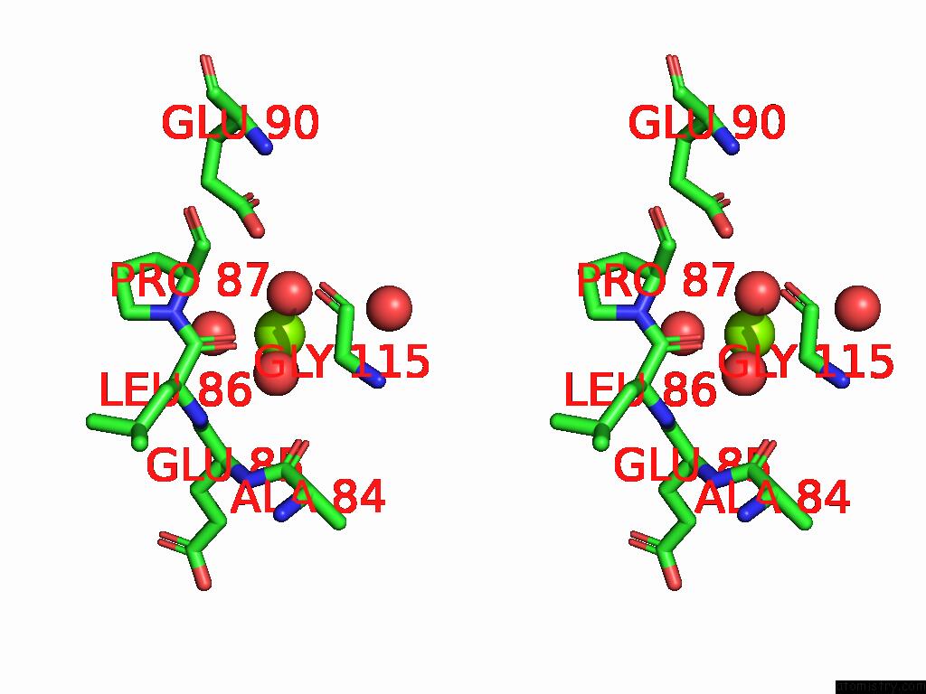
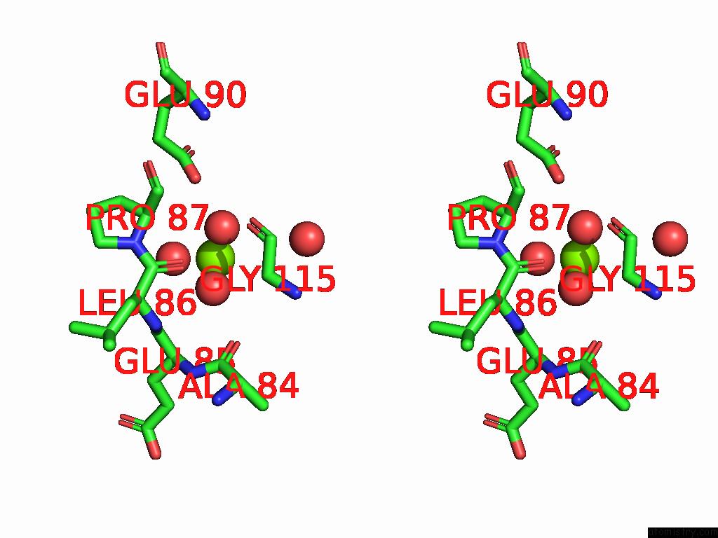
Magnesium binding site 1 out of 5 in 9pg6
Go back to
Magnesium binding site 1 out
of 5 in the Crystal Structure of Glyceraldehyde-3-Phosphate Dehydrogenase From Bordetella Pertussis (Nad Bound)
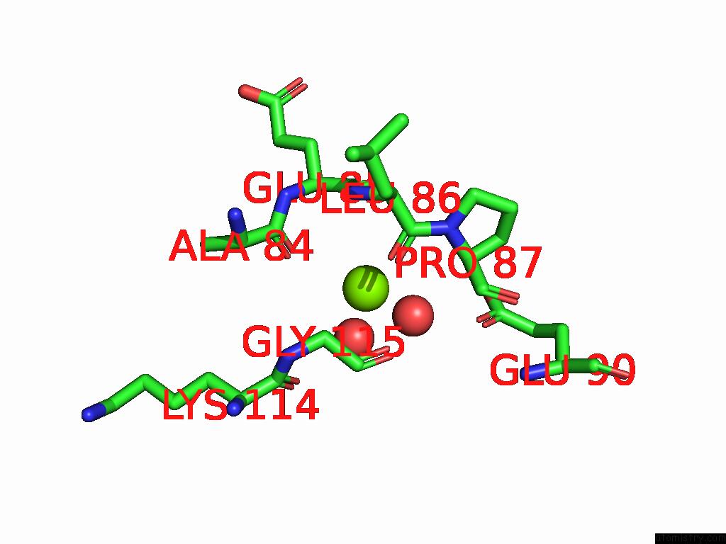
Mono view
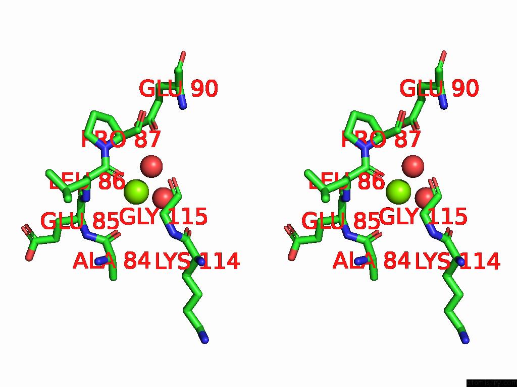
Stereo pair view
Mono view
Stereo pair view
A full contact list of Magnesium with other atoms in the Mg binding
site number 1 of Crystal Structure of Glyceraldehyde-3-Phosphate Dehydrogenase From Bordetella Pertussis (Nad Bound) within 5.0Å range:
|
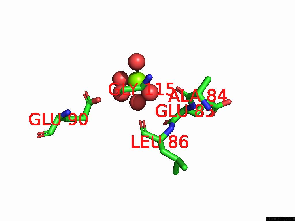
Magnesium binding site 2 out of 5 in 9pg6
Go back to
Magnesium binding site 2 out
of 5 in the Crystal Structure of Glyceraldehyde-3-Phosphate Dehydrogenase From Bordetella Pertussis (Nad Bound)
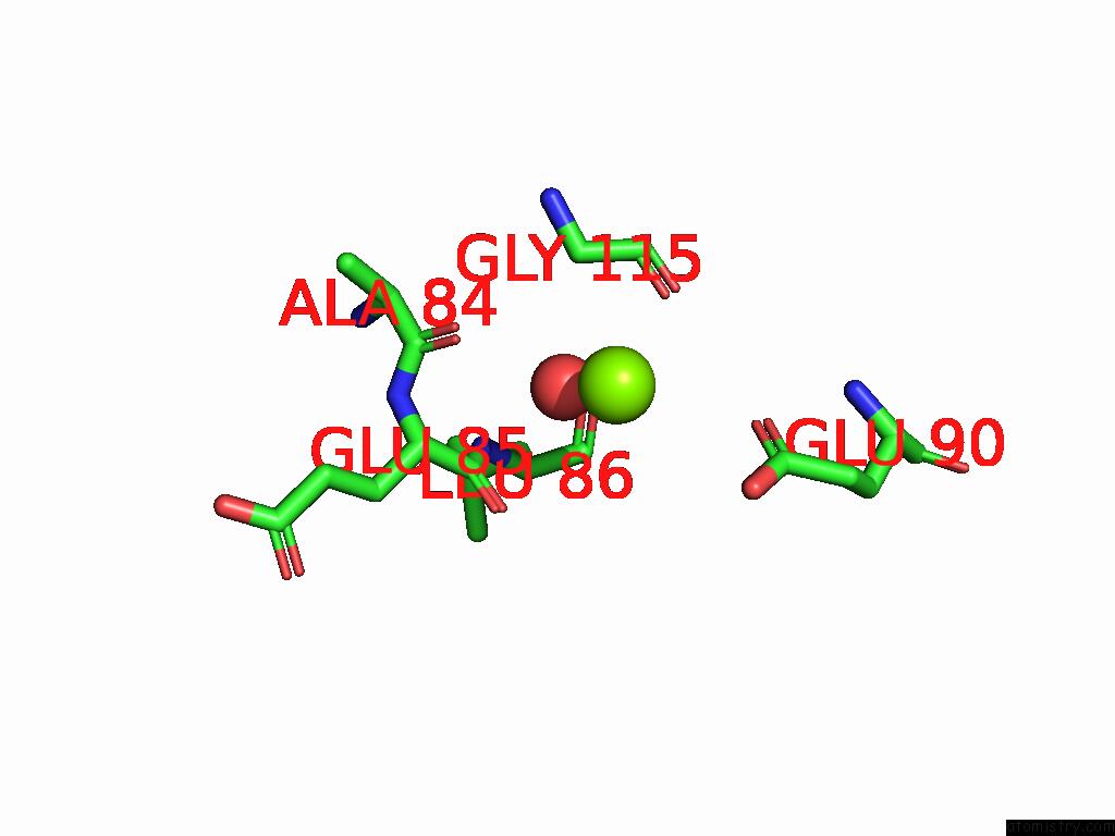
Mono view
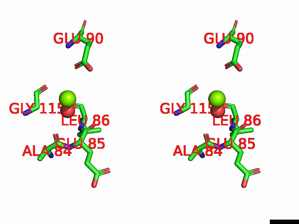
Stereo pair view
Mono view
Stereo pair view
A full contact list of Magnesium with other atoms in the Mg binding
site number 2 of Crystal Structure of Glyceraldehyde-3-Phosphate Dehydrogenase From Bordetella Pertussis (Nad Bound) within 5.0Å range:
|
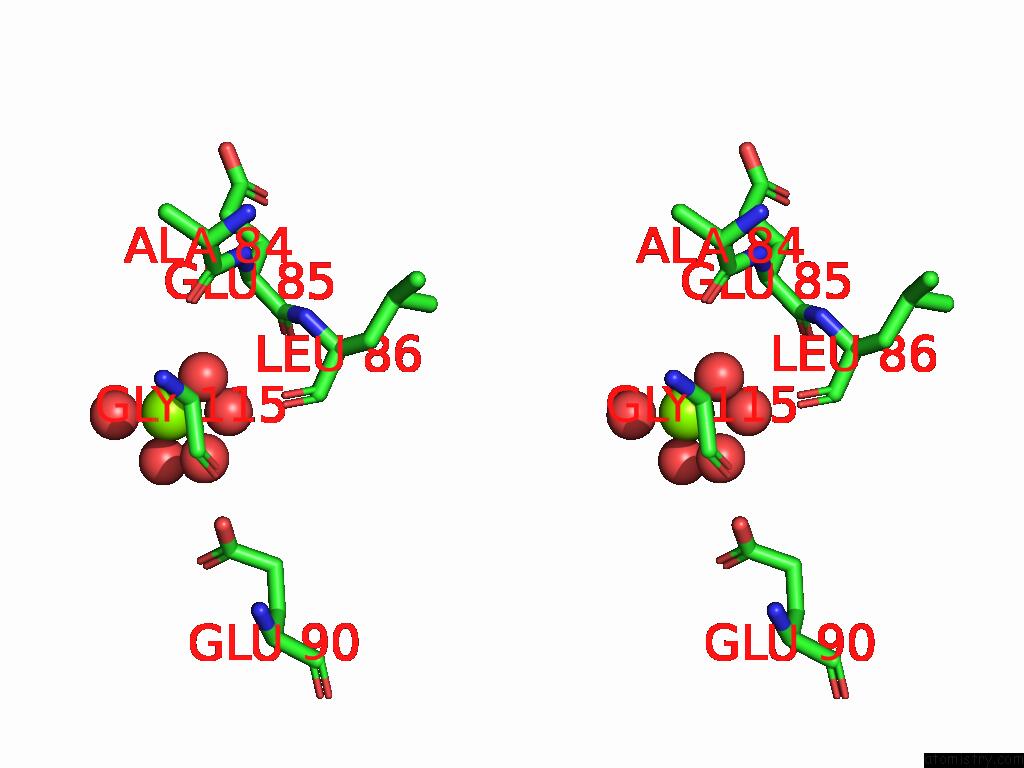
Magnesium binding site 3 out of 5 in 9pg6
Go back to
Magnesium binding site 3 out
of 5 in the Crystal Structure of Glyceraldehyde-3-Phosphate Dehydrogenase From Bordetella Pertussis (Nad Bound)

Mono view
Stereo pair view
Mono view
Stereo pair view
A full contact list of Magnesium with other atoms in the Mg binding
site number 3 of Crystal Structure of Glyceraldehyde-3-Phosphate Dehydrogenase From Bordetella Pertussis (Nad Bound) within 5.0Å range:
|
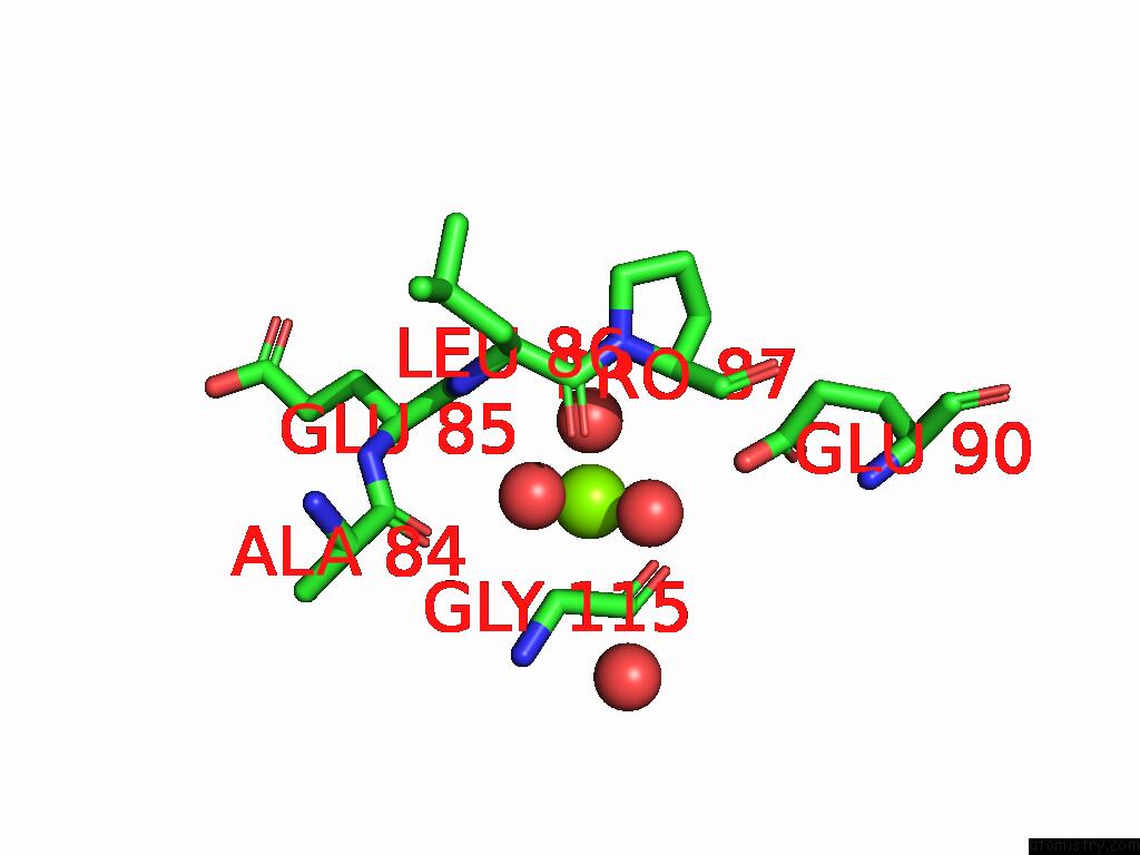
Magnesium binding site 4 out of 5 in 9pg6
Go back to
Magnesium binding site 4 out
of 5 in the Crystal Structure of Glyceraldehyde-3-Phosphate Dehydrogenase From Bordetella Pertussis (Nad Bound)
Mono view
Stereo pair view
Mono view
Stereo pair view
A full contact list of Magnesium with other atoms in the Mg binding
site number 4 of Crystal Structure of Glyceraldehyde-3-Phosphate Dehydrogenase From Bordetella Pertussis (Nad Bound) within 5.0Å range:
|
Magnesium binding site 5 out of 5 in 9pg6
Go back to
Magnesium binding site 5 out
of 5 in the Crystal Structure of Glyceraldehyde-3-Phosphate Dehydrogenase From Bordetella Pertussis (Nad Bound)
Mono view
Stereo pair view
Mono view
Stereo pair view
A full contact list of Magnesium with other atoms in the Mg binding
site number 5 of Crystal Structure of Glyceraldehyde-3-Phosphate Dehydrogenase From Bordetella Pertussis (Nad Bound) within 5.0Å range:
|
Reference:
L.Liu,
A.R.Ung,
S.Lovell,
K.P.Battaile.
Crystal Structure of Glyceraldehyde-3-Phosphate Dehydrogenase From Bordetella Pertussis (Nad Bound) To Be Published.
Page generated: Sat Aug 16 07:10:21 2025
Last articles
Na in 3C2KNa in 3C1Q
Na in 3BPX
Na in 3C0W
Na in 3C0V
Na in 3C0S
Na in 3BX2
Na in 3BZL
Na in 3BVB
Na in 3BV9