Magnesium »
PDB 9nf0-9qof »
9pgq »
Magnesium in PDB 9pgq: Crystal Structure of Glyceraldehyde-3-Phosphate Dehydrogenase From Bordetella Pertussis (Apo, Trigonal Form)
Protein crystallography data
The structure of Crystal Structure of Glyceraldehyde-3-Phosphate Dehydrogenase From Bordetella Pertussis (Apo, Trigonal Form), PDB code: 9pgq
was solved by
Seattle Structural Genomics Center For Infectious Disease,
Seattlestructural Genomics Center For Infectious Disease (Ssgcid),
with X-Ray Crystallography technique. A brief refinement statistics is given in the table below:
| Resolution Low / High (Å) | 43.97 / 1.50 |
| Space group | P 31 2 1 |
| Cell size a, b, c (Å), α, β, γ (°) | 83.461, 83.461, 166.173, 90, 90, 120 |
| R / Rfree (%) | 16.3 / 18.6 |
Magnesium Binding Sites:
The binding sites of Magnesium atom in the Crystal Structure of Glyceraldehyde-3-Phosphate Dehydrogenase From Bordetella Pertussis (Apo, Trigonal Form)
(pdb code 9pgq). This binding sites where shown within
5.0 Angstroms radius around Magnesium atom.
In total 3 binding sites of Magnesium where determined in the Crystal Structure of Glyceraldehyde-3-Phosphate Dehydrogenase From Bordetella Pertussis (Apo, Trigonal Form), PDB code: 9pgq:
Jump to Magnesium binding site number: 1; 2; 3;
In total 3 binding sites of Magnesium where determined in the Crystal Structure of Glyceraldehyde-3-Phosphate Dehydrogenase From Bordetella Pertussis (Apo, Trigonal Form), PDB code: 9pgq:
Jump to Magnesium binding site number: 1; 2; 3;
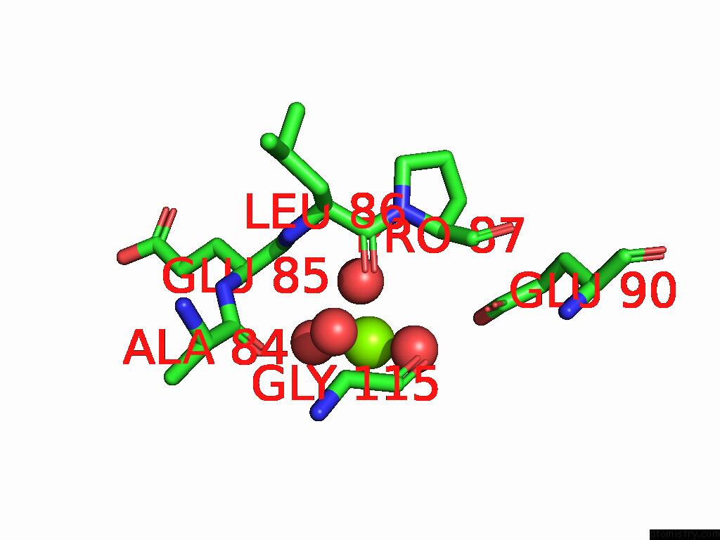
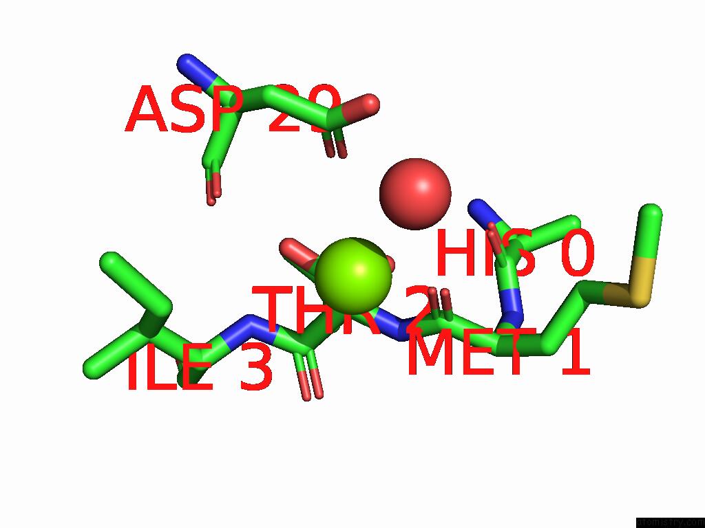
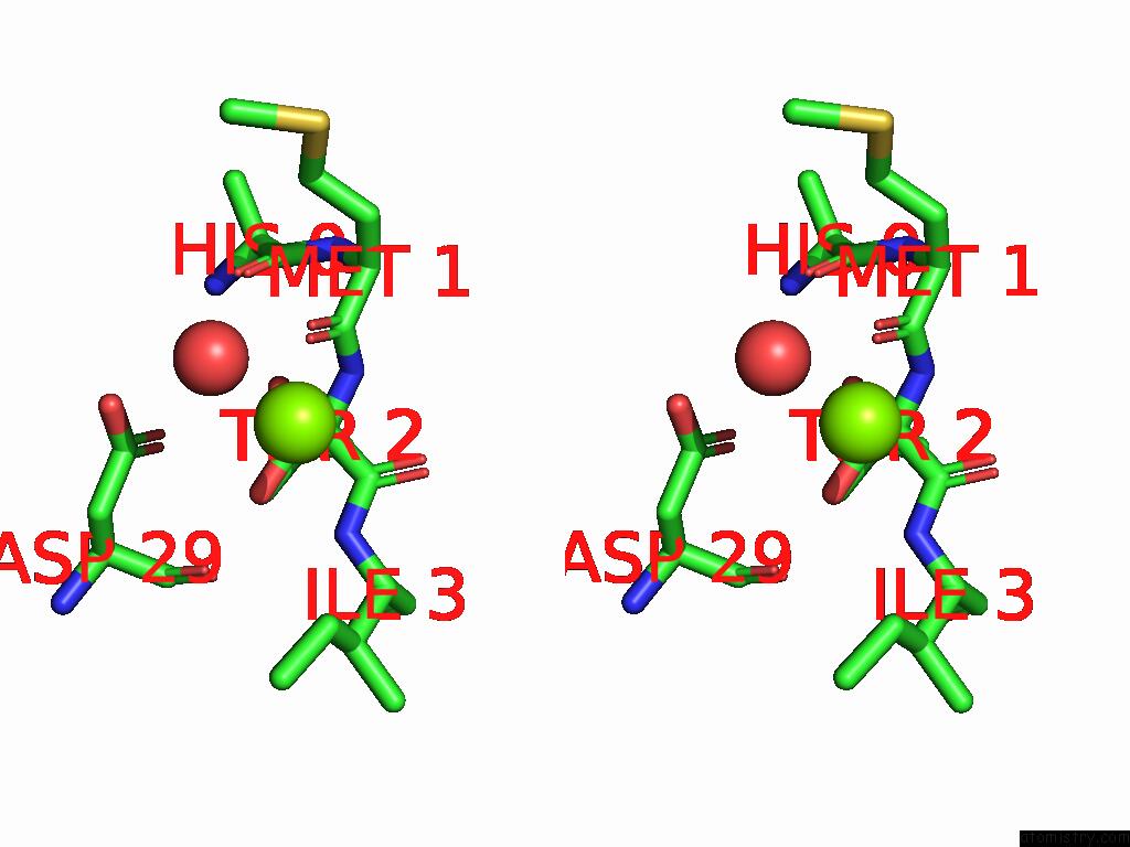
Magnesium binding site 1 out of 3 in 9pgq
Go back to
Magnesium binding site 1 out
of 3 in the Crystal Structure of Glyceraldehyde-3-Phosphate Dehydrogenase From Bordetella Pertussis (Apo, Trigonal Form)
Mono view
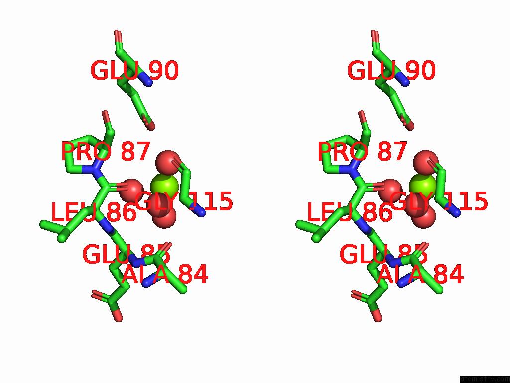
Stereo pair view
Mono view
Stereo pair view
A full contact list of Magnesium with other atoms in the Mg binding
site number 1 of Crystal Structure of Glyceraldehyde-3-Phosphate Dehydrogenase From Bordetella Pertussis (Apo, Trigonal Form) within 5.0Å range:
|
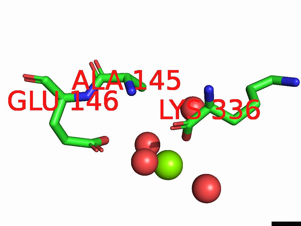
Magnesium binding site 2 out of 3 in 9pgq
Go back to
Magnesium binding site 2 out
of 3 in the Crystal Structure of Glyceraldehyde-3-Phosphate Dehydrogenase From Bordetella Pertussis (Apo, Trigonal Form)
Mono view
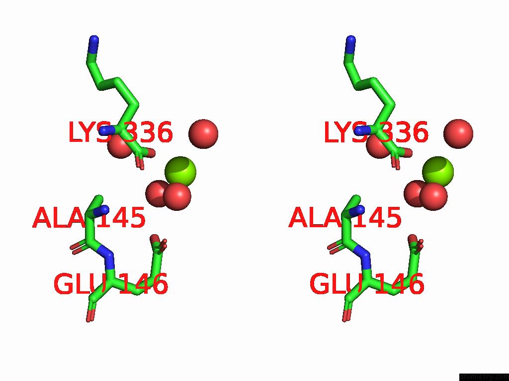
Stereo pair view
Mono view
Stereo pair view
A full contact list of Magnesium with other atoms in the Mg binding
site number 2 of Crystal Structure of Glyceraldehyde-3-Phosphate Dehydrogenase From Bordetella Pertussis (Apo, Trigonal Form) within 5.0Å range:
|
Magnesium binding site 3 out of 3 in 9pgq
Go back to
Magnesium binding site 3 out
of 3 in the Crystal Structure of Glyceraldehyde-3-Phosphate Dehydrogenase From Bordetella Pertussis (Apo, Trigonal Form)
Mono view
Stereo pair view
Mono view
Stereo pair view
A full contact list of Magnesium with other atoms in the Mg binding
site number 3 of Crystal Structure of Glyceraldehyde-3-Phosphate Dehydrogenase From Bordetella Pertussis (Apo, Trigonal Form) within 5.0Å range:
|
Reference:
A.R.Ung,
L.Liu,
S.Lovell,
K.P.Battaile.
Crystal Structure of Glyceraldehyde-3-Phosphate Dehydrogenase From Bordetella Pertussis (Apo, Trigonal Form) To Be Published.
Page generated: Sat Aug 16 07:10:20 2025
Last articles
Na in 6BHDNa in 6BGK
Na in 6BG0
Na in 6BGQ
Na in 6BG1
Na in 6BAQ
Na in 6BFO
Na in 6BFK
Na in 6BEM
Na in 6BEL