Magnesium »
PDB 1fp6-1g7t »
1g7t »
Magnesium in PDB 1g7t: X-Ray Structure of Translation Initiation Factor IF2/EIF5B Complexed with Gdpnp
Protein crystallography data
The structure of X-Ray Structure of Translation Initiation Factor IF2/EIF5B Complexed with Gdpnp, PDB code: 1g7t
was solved by
A.Roll-Mecak,
C.Cao,
T.E.Dever,
S.K.Burley,
with X-Ray Crystallography technique. A brief refinement statistics is given in the table below:
| Resolution Low / High (Å) | 22.00 / 2.00 |
| Space group | P 1 |
| Cell size a, b, c (Å), α, β, γ (°) | 48.799, 53.680, 94.926, 105.17, 94.76, 100.17 |
| R / Rfree (%) | 23.1 / 26.7 |
Magnesium Binding Sites:
The binding sites of Magnesium atom in the X-Ray Structure of Translation Initiation Factor IF2/EIF5B Complexed with Gdpnp
(pdb code 1g7t). This binding sites where shown within
5.0 Angstroms radius around Magnesium atom.
In total only one binding site of Magnesium was determined in the X-Ray Structure of Translation Initiation Factor IF2/EIF5B Complexed with Gdpnp, PDB code: 1g7t:
In total only one binding site of Magnesium was determined in the X-Ray Structure of Translation Initiation Factor IF2/EIF5B Complexed with Gdpnp, PDB code: 1g7t:
Magnesium binding site 1 out of 1 in 1g7t
Go back to
Magnesium binding site 1 out
of 1 in the X-Ray Structure of Translation Initiation Factor IF2/EIF5B Complexed with Gdpnp
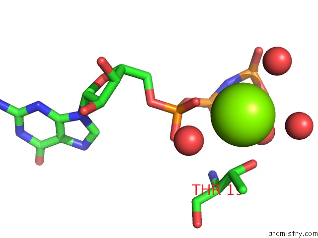
Mono view
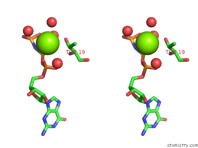
Stereo pair view
Mono view
Stereo pair view
A full contact list of Magnesium with other atoms in the Mg binding
site number 1 of X-Ray Structure of Translation Initiation Factor IF2/EIF5B Complexed with Gdpnp within 5.0Å range:
|
Reference:
A.Roll-Mecak,
C.Cao,
T.E.Dever,
S.K.Burley.
X-Ray Structures of the Universal Translation Initiation Factor IF2/EIF5B: Conformational Changes on Gdp and Gtp Binding. Cell(Cambridge,Mass.) V. 103 781 2000.
ISSN: ISSN 0092-8674
PubMed: 11114334
DOI: 10.1016/S0092-8674(00)00181-1
Page generated: Sat Aug 9 21:16:20 2025
ISSN: ISSN 0092-8674
PubMed: 11114334
DOI: 10.1016/S0092-8674(00)00181-1
Last articles
Mg in 1KJIMg in 1KJ9
Mg in 1KJ8
Mg in 1KIZ
Mg in 1KIL
Mg in 1KHZ
Mg in 1KHK
Mg in 1KH7
Mg in 1KH9
Mg in 1KH5