Magnesium »
PDB 1iah-1iru »
1iir »
Magnesium in PDB 1iir: Crystal Structure of Udp-Glucosyltransferase Gtfb
Protein crystallography data
The structure of Crystal Structure of Udp-Glucosyltransferase Gtfb, PDB code: 1iir
was solved by
A.M.Mulichak,
H.C.Losey,
C.T.Walsh,
R.M.Garavito,
with X-Ray Crystallography technique. A brief refinement statistics is given in the table below:
| Resolution Low / High (Å) | 30.00 / 1.80 |
| Space group | P 41 21 2 |
| Cell size a, b, c (Å), α, β, γ (°) | 102.110, 102.110, 83.350, 90.00, 90.00, 90.00 |
| R / Rfree (%) | 21.1 / 23.1 |
Magnesium Binding Sites:
The binding sites of Magnesium atom in the Crystal Structure of Udp-Glucosyltransferase Gtfb
(pdb code 1iir). This binding sites where shown within
5.0 Angstroms radius around Magnesium atom.
In total 2 binding sites of Magnesium where determined in the Crystal Structure of Udp-Glucosyltransferase Gtfb, PDB code: 1iir:
Jump to Magnesium binding site number: 1; 2;
In total 2 binding sites of Magnesium where determined in the Crystal Structure of Udp-Glucosyltransferase Gtfb, PDB code: 1iir:
Jump to Magnesium binding site number: 1; 2;
Magnesium binding site 1 out of 2 in 1iir
Go back to
Magnesium binding site 1 out
of 2 in the Crystal Structure of Udp-Glucosyltransferase Gtfb

Mono view
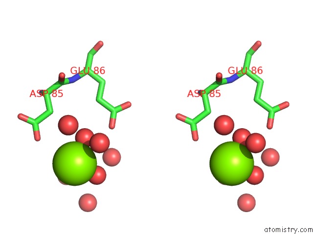
Stereo pair view
Mono view
Stereo pair view
A full contact list of Magnesium with other atoms in the Mg binding
site number 1 of Crystal Structure of Udp-Glucosyltransferase Gtfb within 5.0Å range:
|
Magnesium binding site 2 out of 2 in 1iir
Go back to
Magnesium binding site 2 out
of 2 in the Crystal Structure of Udp-Glucosyltransferase Gtfb
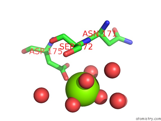
Mono view
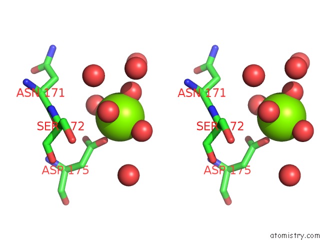
Stereo pair view
Mono view
Stereo pair view
A full contact list of Magnesium with other atoms in the Mg binding
site number 2 of Crystal Structure of Udp-Glucosyltransferase Gtfb within 5.0Å range:
|
Reference:
A.M.Mulichak,
H.C.Losey,
C.T.Walsh,
R.M.Garavito.
Structure of the Udp-Glucosyltransferase Gtfb That Modifies the Heptapeptide Aglycone in the Biosynthesis of Vancomycin Group Antibiotics. Structure V. 9 547 2001.
ISSN: ISSN 0969-2126
PubMed: 11470430
DOI: 10.1016/S0969-2126(01)00616-5
Page generated: Sat Aug 9 22:19:42 2025
ISSN: ISSN 0969-2126
PubMed: 11470430
DOI: 10.1016/S0969-2126(01)00616-5
Last articles
Mg in 1SKSMg in 1SJC
Mg in 1SJB
Mg in 1SKQ
Mg in 1SJA
Mg in 1SJN
Mg in 1SJ3
Mg in 1SIJ
Mg in 1SIX
Mg in 1SHZ