Magnesium »
PDB 1iah-1iru »
1iru »
Magnesium in PDB 1iru: Crystal Structure of the Mammalian 20S Proteasome at 2.75 A Resolution
Protein crystallography data
The structure of Crystal Structure of the Mammalian 20S Proteasome at 2.75 A Resolution, PDB code: 1iru
was solved by
M.Unno,
T.Mizushima,
Y.Morimoto,
Y.Tomisugi,
K.Tanaka,
N.Yasuoka,
T.Tsukihara,
with X-Ray Crystallography technique. A brief refinement statistics is given in the table below:
| Resolution Low / High (Å) | 65.00 / 2.75 |
| Space group | P 21 21 21 |
| Cell size a, b, c (Å), α, β, γ (°) | 316.700, 205.900, 116.000, 90.00, 90.00, 90.00 |
| R / Rfree (%) | 25 / 29.4 |
Magnesium Binding Sites:
Pages:
>>> Page 1 <<< Page 2, Binding sites: 11 - 20; Page 3, Binding sites: 21 - 30;Binding sites:
The binding sites of Magnesium atom in the Crystal Structure of the Mammalian 20S Proteasome at 2.75 A Resolution (pdb code 1iru). This binding sites where shown within 5.0 Angstroms radius around Magnesium atom.In total 30 binding sites of Magnesium where determined in the Crystal Structure of the Mammalian 20S Proteasome at 2.75 A Resolution, PDB code: 1iru:
Jump to Magnesium binding site number: 1; 2; 3; 4; 5; 6; 7; 8; 9; 10;
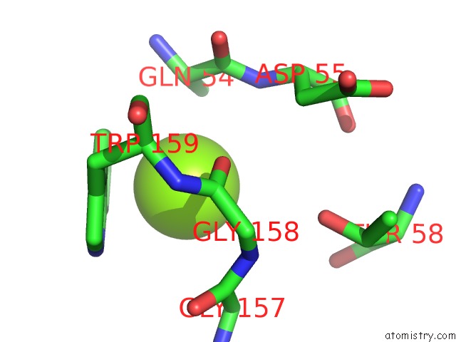
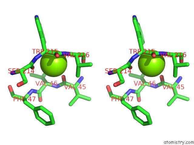
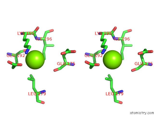
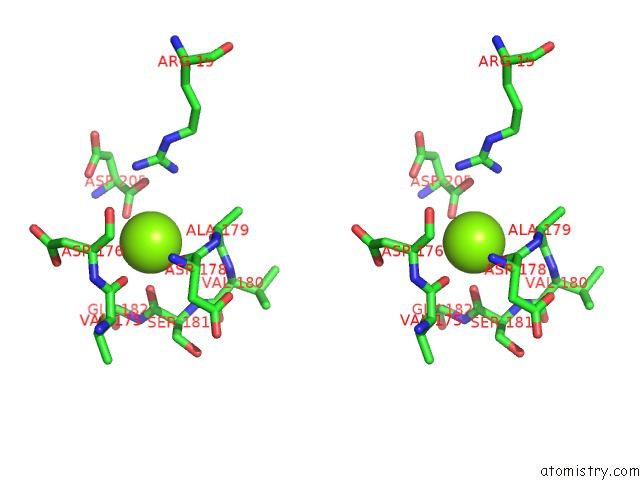
Magnesium binding site 1 out of 30 in 1iru
Go back to
Magnesium binding site 1 out
of 30 in the Crystal Structure of the Mammalian 20S Proteasome at 2.75 A Resolution
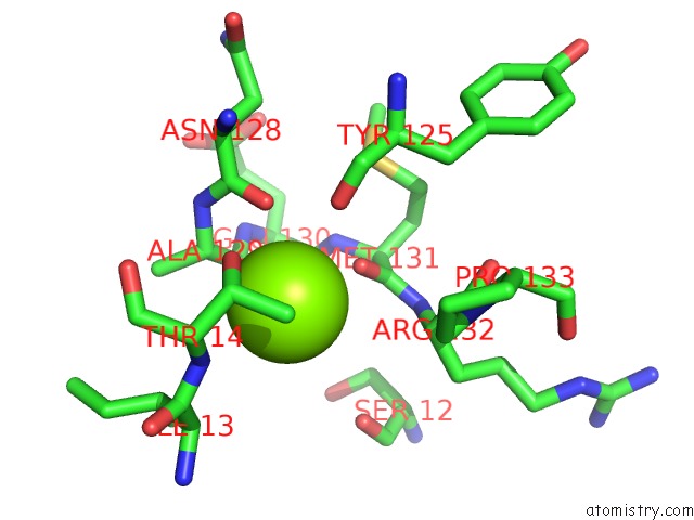
Mono view
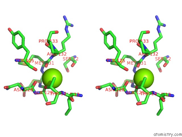
Stereo pair view
Mono view
Stereo pair view
A full contact list of Magnesium with other atoms in the Mg binding
site number 1 of Crystal Structure of the Mammalian 20S Proteasome at 2.75 A Resolution within 5.0Å range:
|
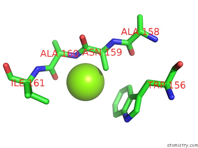
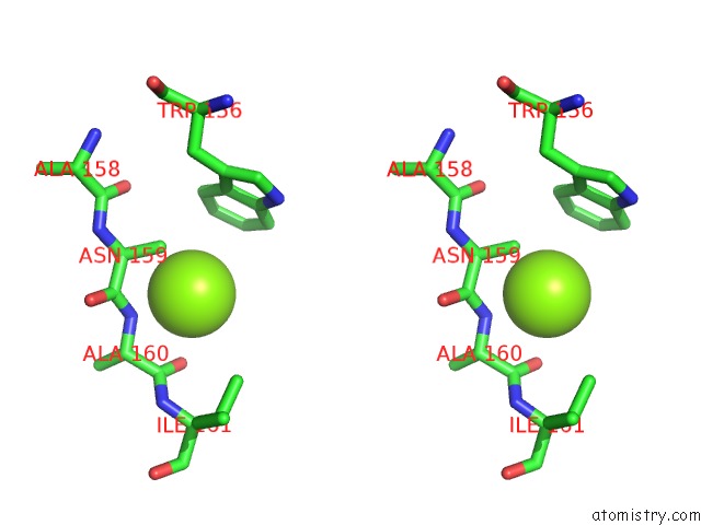
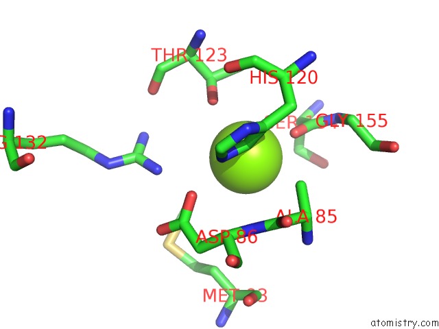
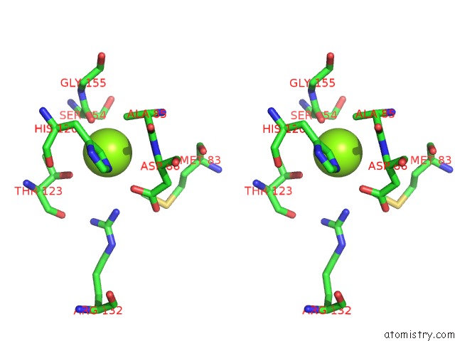
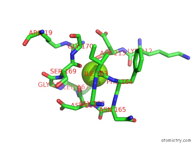
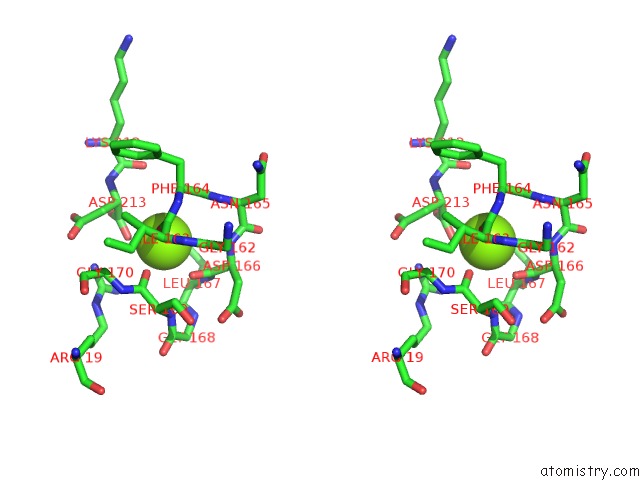
Magnesium binding site 2 out of 30 in 1iru
Go back to
Magnesium binding site 2 out
of 30 in the Crystal Structure of the Mammalian 20S Proteasome at 2.75 A Resolution
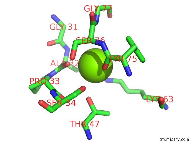
Mono view
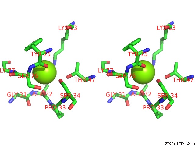
Stereo pair view
Mono view
Stereo pair view
A full contact list of Magnesium with other atoms in the Mg binding
site number 2 of Crystal Structure of the Mammalian 20S Proteasome at 2.75 A Resolution within 5.0Å range:
|
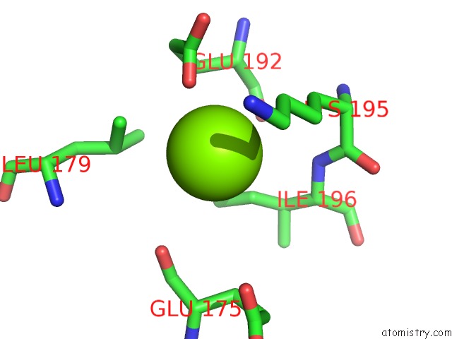
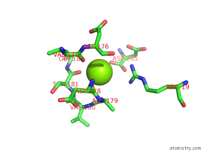
Magnesium binding site 3 out of 30 in 1iru
Go back to
Magnesium binding site 3 out
of 30 in the Crystal Structure of the Mammalian 20S Proteasome at 2.75 A Resolution
Mono view

Stereo pair view
Mono view
Stereo pair view
A full contact list of Magnesium with other atoms in the Mg binding
site number 3 of Crystal Structure of the Mammalian 20S Proteasome at 2.75 A Resolution within 5.0Å range:
|
Magnesium binding site 4 out of 30 in 1iru
Go back to
Magnesium binding site 4 out
of 30 in the Crystal Structure of the Mammalian 20S Proteasome at 2.75 A Resolution
Mono view
Stereo pair view
Mono view
Stereo pair view
A full contact list of Magnesium with other atoms in the Mg binding
site number 4 of Crystal Structure of the Mammalian 20S Proteasome at 2.75 A Resolution within 5.0Å range:
|
Magnesium binding site 5 out of 30 in 1iru
Go back to
Magnesium binding site 5 out
of 30 in the Crystal Structure of the Mammalian 20S Proteasome at 2.75 A Resolution
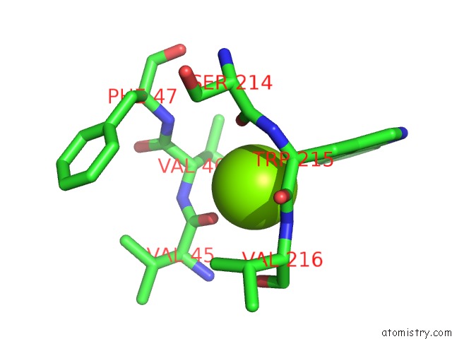
Mono view
Stereo pair view
Mono view
Stereo pair view
A full contact list of Magnesium with other atoms in the Mg binding
site number 5 of Crystal Structure of the Mammalian 20S Proteasome at 2.75 A Resolution within 5.0Å range:
|
Magnesium binding site 6 out of 30 in 1iru
Go back to
Magnesium binding site 6 out
of 30 in the Crystal Structure of the Mammalian 20S Proteasome at 2.75 A Resolution
Mono view
Stereo pair view
Mono view
Stereo pair view
A full contact list of Magnesium with other atoms in the Mg binding
site number 6 of Crystal Structure of the Mammalian 20S Proteasome at 2.75 A Resolution within 5.0Å range:
|
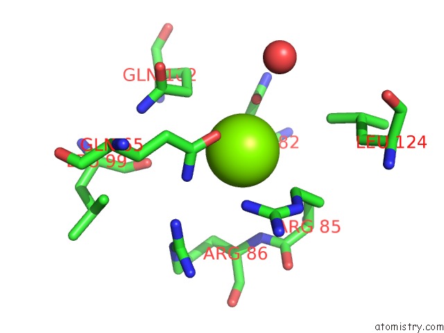
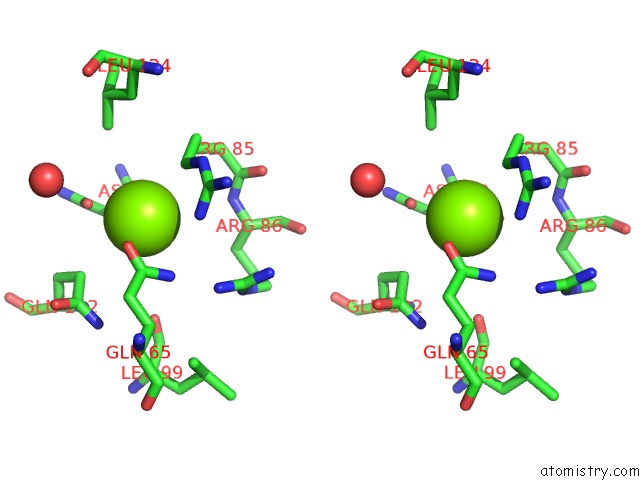
Magnesium binding site 7 out of 30 in 1iru
Go back to
Magnesium binding site 7 out
of 30 in the Crystal Structure of the Mammalian 20S Proteasome at 2.75 A Resolution
Mono view
Stereo pair view
Mono view
Stereo pair view
A full contact list of Magnesium with other atoms in the Mg binding
site number 7 of Crystal Structure of the Mammalian 20S Proteasome at 2.75 A Resolution within 5.0Å range:
|
Magnesium binding site 8 out of 30 in 1iru
Go back to
Magnesium binding site 8 out
of 30 in the Crystal Structure of the Mammalian 20S Proteasome at 2.75 A Resolution
Mono view
Stereo pair view
Mono view
Stereo pair view
A full contact list of Magnesium with other atoms in the Mg binding
site number 8 of Crystal Structure of the Mammalian 20S Proteasome at 2.75 A Resolution within 5.0Å range:
|
Magnesium binding site 9 out of 30 in 1iru
Go back to
Magnesium binding site 9 out
of 30 in the Crystal Structure of the Mammalian 20S Proteasome at 2.75 A Resolution
Mono view
Stereo pair view
Mono view
Stereo pair view
A full contact list of Magnesium with other atoms in the Mg binding
site number 9 of Crystal Structure of the Mammalian 20S Proteasome at 2.75 A Resolution within 5.0Å range:
|
Magnesium binding site 10 out of 30 in 1iru
Go back to
Magnesium binding site 10 out
of 30 in the Crystal Structure of the Mammalian 20S Proteasome at 2.75 A Resolution
Mono view
Stereo pair view
Mono view
Stereo pair view
A full contact list of Magnesium with other atoms in the Mg binding
site number 10 of Crystal Structure of the Mammalian 20S Proteasome at 2.75 A Resolution within 5.0Å range:
|
Reference:
M.Unno,
T.Mizushima,
Y.Morimoto,
Y.Tomisugi,
K.Tanaka,
N.Yasuoka,
T.Tsukihara.
The Structure of the Mammalian 20S Proteasome at 2.75 A Resolution. Structure V. 10 609 2002.
ISSN: ISSN 0969-2126
PubMed: 12015144
DOI: 10.1016/S0969-2126(02)00748-7
Page generated: Sat Aug 9 22:41:43 2025
ISSN: ISSN 0969-2126
PubMed: 12015144
DOI: 10.1016/S0969-2126(02)00748-7
Last articles
Mg in 1SKRMg in 1SL0
Mg in 1SL1
Mg in 1SKW
Mg in 1SKS
Mg in 1SJC
Mg in 1SJB
Mg in 1SKQ
Mg in 1SJA
Mg in 1SJN