Magnesium »
PDB 1it7-1jbz »
1ixj »
Magnesium in PDB 1ixj: Crystal Structure of D(Gcgaaagct) Containing Parallel- Stranded Duplex with Homo Base Pairs and Anti-Parallel Duplex with Watson-Crick Base Pairs
Protein crystallography data
The structure of Crystal Structure of D(Gcgaaagct) Containing Parallel- Stranded Duplex with Homo Base Pairs and Anti-Parallel Duplex with Watson-Crick Base Pairs, PDB code: 1ixj
was solved by
T.Sunami,
J.Kondo,
T.Kobuna,
I.Hirao,
K.Watanabe,
K.Miura,
A.Takenaka,
with X-Ray Crystallography technique. A brief refinement statistics is given in the table below:
| Resolution Low / High (Å) | 9.00 / 2.50 |
| Space group | I 41 2 2 |
| Cell size a, b, c (Å), α, β, γ (°) | 53.400, 53.400, 54.000, 90.00, 90.00, 90.00 |
| R / Rfree (%) | 21.5 / 25.7 |
Other elements in 1ixj:
The structure of Crystal Structure of D(Gcgaaagct) Containing Parallel- Stranded Duplex with Homo Base Pairs and Anti-Parallel Duplex with Watson-Crick Base Pairs also contains other interesting chemical elements:
| Cobalt | (Co) | 1 atom |
Magnesium Binding Sites:
The binding sites of Magnesium atom in the Crystal Structure of D(Gcgaaagct) Containing Parallel- Stranded Duplex with Homo Base Pairs and Anti-Parallel Duplex with Watson-Crick Base Pairs
(pdb code 1ixj). This binding sites where shown within
5.0 Angstroms radius around Magnesium atom.
In total 2 binding sites of Magnesium where determined in the Crystal Structure of D(Gcgaaagct) Containing Parallel- Stranded Duplex with Homo Base Pairs and Anti-Parallel Duplex with Watson-Crick Base Pairs, PDB code: 1ixj:
Jump to Magnesium binding site number: 1; 2;
In total 2 binding sites of Magnesium where determined in the Crystal Structure of D(Gcgaaagct) Containing Parallel- Stranded Duplex with Homo Base Pairs and Anti-Parallel Duplex with Watson-Crick Base Pairs, PDB code: 1ixj:
Jump to Magnesium binding site number: 1; 2;
Magnesium binding site 1 out of 2 in 1ixj
Go back to
Magnesium binding site 1 out
of 2 in the Crystal Structure of D(Gcgaaagct) Containing Parallel- Stranded Duplex with Homo Base Pairs and Anti-Parallel Duplex with Watson-Crick Base Pairs
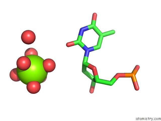
Mono view
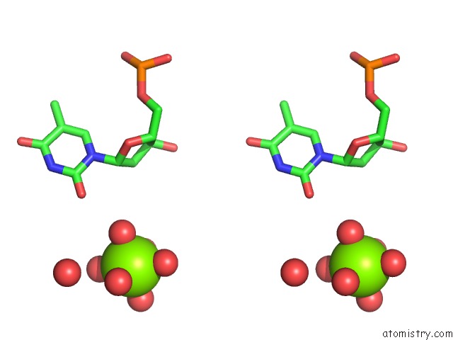
Stereo pair view
Mono view
Stereo pair view
A full contact list of Magnesium with other atoms in the Mg binding
site number 1 of Crystal Structure of D(Gcgaaagct) Containing Parallel- Stranded Duplex with Homo Base Pairs and Anti-Parallel Duplex with Watson-Crick Base Pairs within 5.0Å range:
|
Magnesium binding site 2 out of 2 in 1ixj
Go back to
Magnesium binding site 2 out
of 2 in the Crystal Structure of D(Gcgaaagct) Containing Parallel- Stranded Duplex with Homo Base Pairs and Anti-Parallel Duplex with Watson-Crick Base Pairs
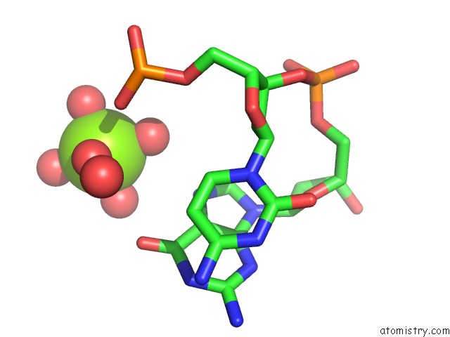
Mono view
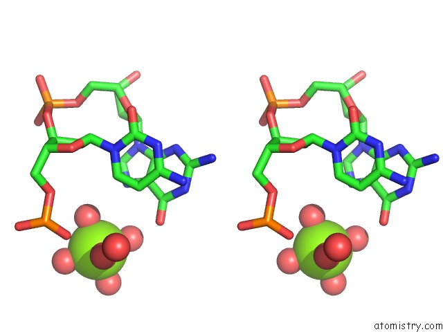
Stereo pair view
Mono view
Stereo pair view
A full contact list of Magnesium with other atoms in the Mg binding
site number 2 of Crystal Structure of D(Gcgaaagct) Containing Parallel- Stranded Duplex with Homo Base Pairs and Anti-Parallel Duplex with Watson-Crick Base Pairs within 5.0Å range:
|
Reference:
T.Sunami,
J.Kondo,
T.Kobuna,
I.Hirao,
K.Watanabe,
K.Miura,
A.Takenaka.
Crystal Structure of D(Gcgaaagct) Containing A Parallel-Stranded Duplex with Homo Base Pairs and An Anti-Parallel Duplex with Watson-Crick Base Pairs Nucleic Acids Res. V. 30 5253 2002.
ISSN: ISSN 0305-1048
PubMed: 12466550
DOI: 10.1093/NAR/GKF639
Page generated: Sat Aug 9 22:47:24 2025
ISSN: ISSN 0305-1048
PubMed: 12466550
DOI: 10.1093/NAR/GKF639
Last articles
Mg in 2CHGMg in 2CIC
Mg in 2CHM
Mg in 2CEA
Mg in 2CE7
Mg in 2CHE
Mg in 2CG5
Mg in 2CFS
Mg in 2CG4
Mg in 2CFR