Magnesium »
PDB 1l3r-1lny »
1l6s »
Magnesium in PDB 1l6s: Crystal Structure of Porphobilinogen Synthase Complexed with the Inhibitor 4,7-Dioxosebacic Acid
Enzymatic activity of Crystal Structure of Porphobilinogen Synthase Complexed with the Inhibitor 4,7-Dioxosebacic Acid
All present enzymatic activity of Crystal Structure of Porphobilinogen Synthase Complexed with the Inhibitor 4,7-Dioxosebacic Acid:
4.2.1.24;
4.2.1.24;
Protein crystallography data
The structure of Crystal Structure of Porphobilinogen Synthase Complexed with the Inhibitor 4,7-Dioxosebacic Acid, PDB code: 1l6s
was solved by
E.K.Jaffe,
J.Kervinen,
J.Martins,
F.Stauffer,
R.Neier,
A.Wlodawer,
A.Zdanov,
with X-Ray Crystallography technique. A brief refinement statistics is given in the table below:
| Resolution Low / High (Å) | 40.00 / 1.70 |
| Space group | P 4 21 2 |
| Cell size a, b, c (Å), α, β, γ (°) | 129.000, 129.000, 142.800, 90.00, 90.00, 90.00 |
| R / Rfree (%) | 19.5 / 24.3 |
Other elements in 1l6s:
The structure of Crystal Structure of Porphobilinogen Synthase Complexed with the Inhibitor 4,7-Dioxosebacic Acid also contains other interesting chemical elements:
| Zinc | (Zn) | 2 atoms |
Magnesium Binding Sites:
The binding sites of Magnesium atom in the Crystal Structure of Porphobilinogen Synthase Complexed with the Inhibitor 4,7-Dioxosebacic Acid
(pdb code 1l6s). This binding sites where shown within
5.0 Angstroms radius around Magnesium atom.
In total 2 binding sites of Magnesium where determined in the Crystal Structure of Porphobilinogen Synthase Complexed with the Inhibitor 4,7-Dioxosebacic Acid, PDB code: 1l6s:
Jump to Magnesium binding site number: 1; 2;
In total 2 binding sites of Magnesium where determined in the Crystal Structure of Porphobilinogen Synthase Complexed with the Inhibitor 4,7-Dioxosebacic Acid, PDB code: 1l6s:
Jump to Magnesium binding site number: 1; 2;
Magnesium binding site 1 out of 2 in 1l6s
Go back to
Magnesium binding site 1 out
of 2 in the Crystal Structure of Porphobilinogen Synthase Complexed with the Inhibitor 4,7-Dioxosebacic Acid
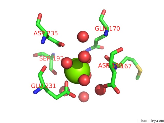
Mono view
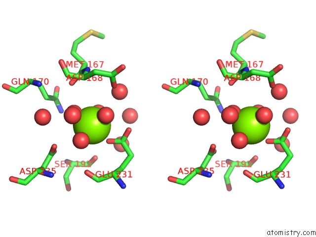
Stereo pair view
Mono view
Stereo pair view
A full contact list of Magnesium with other atoms in the Mg binding
site number 1 of Crystal Structure of Porphobilinogen Synthase Complexed with the Inhibitor 4,7-Dioxosebacic Acid within 5.0Å range:
|
Magnesium binding site 2 out of 2 in 1l6s
Go back to
Magnesium binding site 2 out
of 2 in the Crystal Structure of Porphobilinogen Synthase Complexed with the Inhibitor 4,7-Dioxosebacic Acid
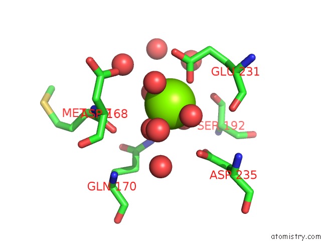
Mono view
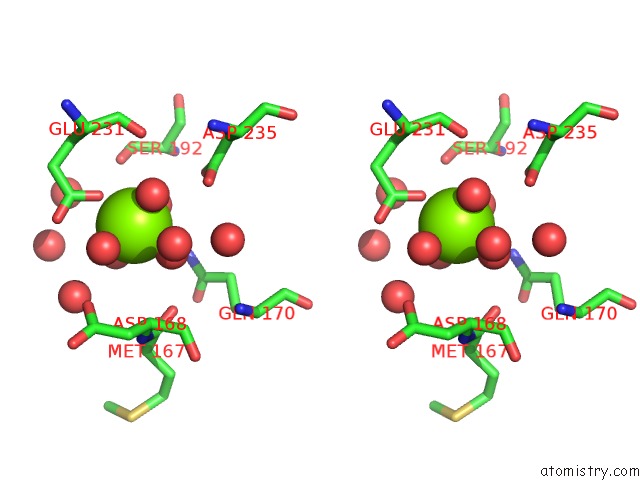
Stereo pair view
Mono view
Stereo pair view
A full contact list of Magnesium with other atoms in the Mg binding
site number 2 of Crystal Structure of Porphobilinogen Synthase Complexed with the Inhibitor 4,7-Dioxosebacic Acid within 5.0Å range:
|
Reference:
E.K.Jaffe,
J.Kervinen,
J.Martins,
F.Stauffer,
R.Neier,
A.Wlodawer,
A.Zdanov.
Species-Specific Inhibition of Porphobilinogen Synthase By 4-Oxosebacic Acid J.Biol.Chem. V. 277 19792 2002.
ISSN: ISSN 0021-9258
PubMed: 11909869
DOI: 10.1074/JBC.M201486200
Page generated: Sun Aug 10 00:38:04 2025
ISSN: ISSN 0021-9258
PubMed: 11909869
DOI: 10.1074/JBC.M201486200
Last articles
Mg in 6TR4Mg in 6TR3
Mg in 6TMF
Mg in 6TQO
Mg in 6TQN
Mg in 6TQF
Mg in 6TQE
Mg in 6TQB
Mg in 6TQA
Mg in 6TPS