Magnesium »
PDB 1l3r-1lny »
1l9j »
Magnesium in PDB 1l9j: X-Ray Structure of the Cytochrome-C(2)-Photosynthetic Reaction Center Electron Transfer Complex From Rhodobacter Sphaeroides in Type I Co- Crystals
Protein crystallography data
The structure of X-Ray Structure of the Cytochrome-C(2)-Photosynthetic Reaction Center Electron Transfer Complex From Rhodobacter Sphaeroides in Type I Co- Crystals, PDB code: 1l9j
was solved by
H.L.Axelrod,
E.C.Abresch,
M.Y.Okamura,
A.P.Yeh,
D.C.Rees,
G.Feher,
with X-Ray Crystallography technique. A brief refinement statistics is given in the table below:
| Resolution Low / High (Å) | 49.32 / 3.25 |
| Space group | P 1 21 1 |
| Cell size a, b, c (Å), α, β, γ (°) | 77.930, 80.312, 246.575, 90.00, 92.41, 90.00 |
| R / Rfree (%) | 24.8 / 28.7 |
Other elements in 1l9j:
The structure of X-Ray Structure of the Cytochrome-C(2)-Photosynthetic Reaction Center Electron Transfer Complex From Rhodobacter Sphaeroides in Type I Co- Crystals also contains other interesting chemical elements:
| Iron | (Fe) | 4 atoms |
| Chlorine | (Cl) | 2 atoms |
Magnesium Binding Sites:
The binding sites of Magnesium atom in the X-Ray Structure of the Cytochrome-C(2)-Photosynthetic Reaction Center Electron Transfer Complex From Rhodobacter Sphaeroides in Type I Co- Crystals
(pdb code 1l9j). This binding sites where shown within
5.0 Angstroms radius around Magnesium atom.
In total 8 binding sites of Magnesium where determined in the X-Ray Structure of the Cytochrome-C(2)-Photosynthetic Reaction Center Electron Transfer Complex From Rhodobacter Sphaeroides in Type I Co- Crystals, PDB code: 1l9j:
Jump to Magnesium binding site number: 1; 2; 3; 4; 5; 6; 7; 8;
In total 8 binding sites of Magnesium where determined in the X-Ray Structure of the Cytochrome-C(2)-Photosynthetic Reaction Center Electron Transfer Complex From Rhodobacter Sphaeroides in Type I Co- Crystals, PDB code: 1l9j:
Jump to Magnesium binding site number: 1; 2; 3; 4; 5; 6; 7; 8;
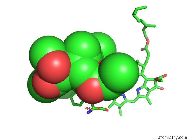
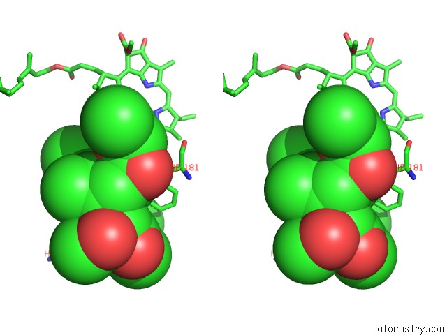
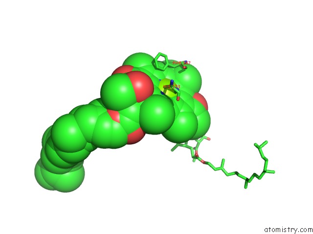
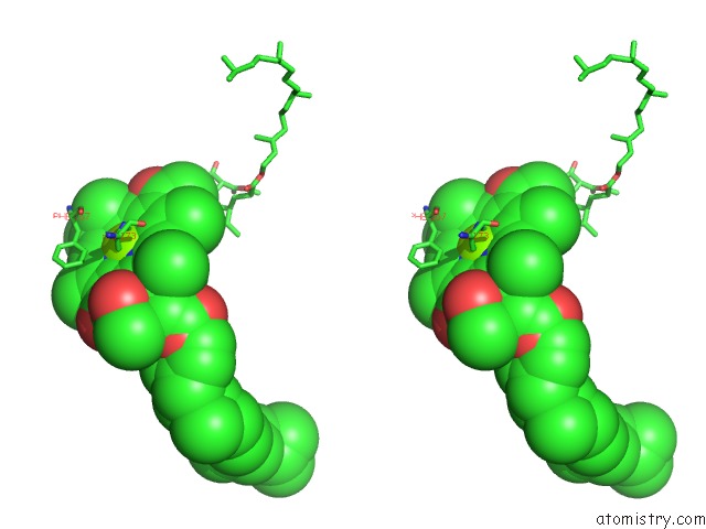
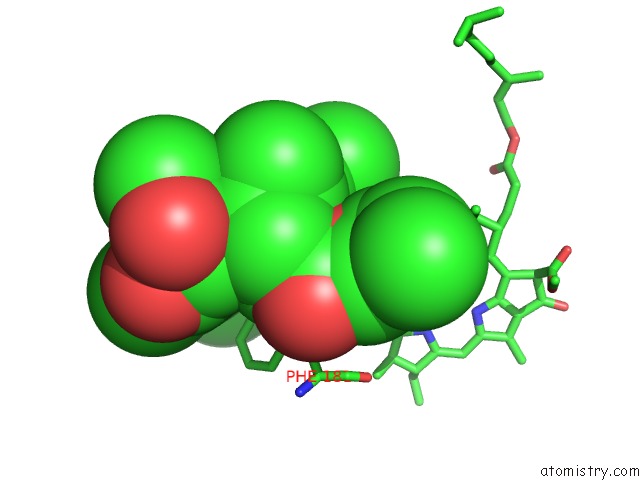
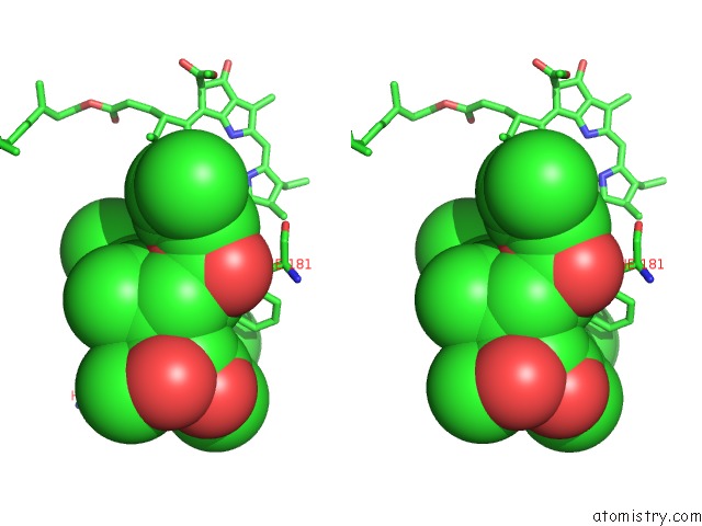
Magnesium binding site 1 out of 8 in 1l9j
Go back to
Magnesium binding site 1 out
of 8 in the X-Ray Structure of the Cytochrome-C(2)-Photosynthetic Reaction Center Electron Transfer Complex From Rhodobacter Sphaeroides in Type I Co- Crystals
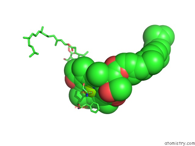
Mono view
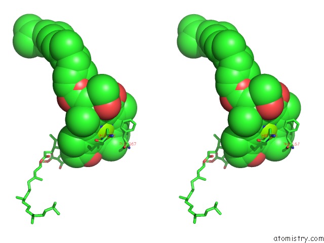
Stereo pair view
Mono view
Stereo pair view
A full contact list of Magnesium with other atoms in the Mg binding
site number 1 of X-Ray Structure of the Cytochrome-C(2)-Photosynthetic Reaction Center Electron Transfer Complex From Rhodobacter Sphaeroides in Type I Co- Crystals within 5.0Å range:
|
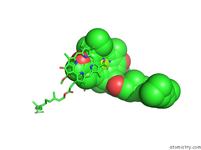
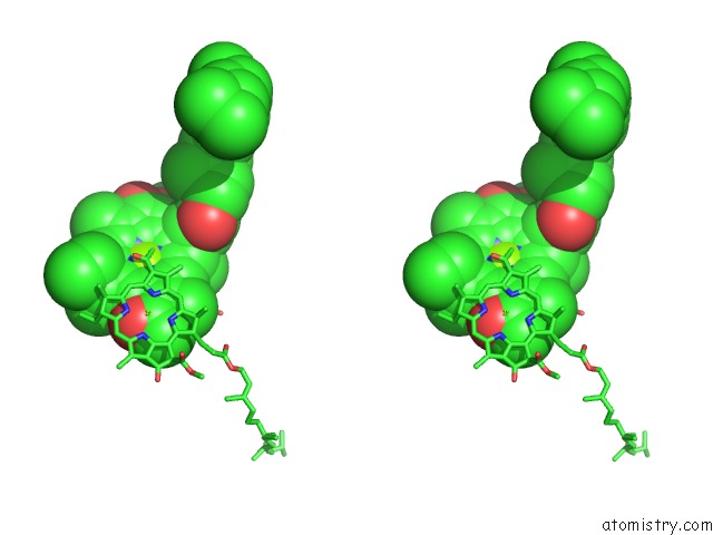
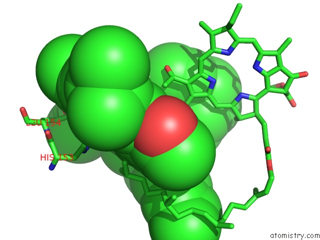
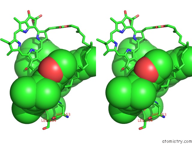
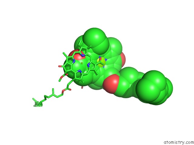
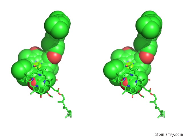
Magnesium binding site 2 out of 8 in 1l9j
Go back to
Magnesium binding site 2 out
of 8 in the X-Ray Structure of the Cytochrome-C(2)-Photosynthetic Reaction Center Electron Transfer Complex From Rhodobacter Sphaeroides in Type I Co- Crystals
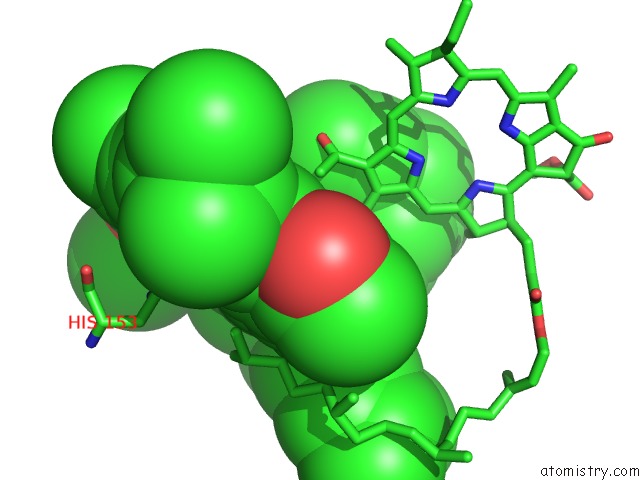
Mono view
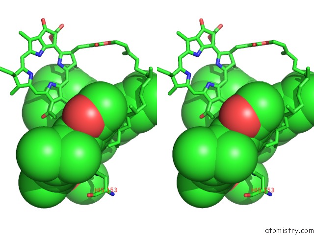
Stereo pair view
Mono view
Stereo pair view
A full contact list of Magnesium with other atoms in the Mg binding
site number 2 of X-Ray Structure of the Cytochrome-C(2)-Photosynthetic Reaction Center Electron Transfer Complex From Rhodobacter Sphaeroides in Type I Co- Crystals within 5.0Å range:
|
Magnesium binding site 3 out of 8 in 1l9j
Go back to
Magnesium binding site 3 out
of 8 in the X-Ray Structure of the Cytochrome-C(2)-Photosynthetic Reaction Center Electron Transfer Complex From Rhodobacter Sphaeroides in Type I Co- Crystals
Mono view
Stereo pair view
Mono view
Stereo pair view
A full contact list of Magnesium with other atoms in the Mg binding
site number 3 of X-Ray Structure of the Cytochrome-C(2)-Photosynthetic Reaction Center Electron Transfer Complex From Rhodobacter Sphaeroides in Type I Co- Crystals within 5.0Å range:
|
Magnesium binding site 4 out of 8 in 1l9j
Go back to
Magnesium binding site 4 out
of 8 in the X-Ray Structure of the Cytochrome-C(2)-Photosynthetic Reaction Center Electron Transfer Complex From Rhodobacter Sphaeroides in Type I Co- Crystals
Mono view
Stereo pair view
Mono view
Stereo pair view
A full contact list of Magnesium with other atoms in the Mg binding
site number 4 of X-Ray Structure of the Cytochrome-C(2)-Photosynthetic Reaction Center Electron Transfer Complex From Rhodobacter Sphaeroides in Type I Co- Crystals within 5.0Å range:
|
Magnesium binding site 5 out of 8 in 1l9j
Go back to
Magnesium binding site 5 out
of 8 in the X-Ray Structure of the Cytochrome-C(2)-Photosynthetic Reaction Center Electron Transfer Complex From Rhodobacter Sphaeroides in Type I Co- Crystals
Mono view
Stereo pair view
Mono view
Stereo pair view
A full contact list of Magnesium with other atoms in the Mg binding
site number 5 of X-Ray Structure of the Cytochrome-C(2)-Photosynthetic Reaction Center Electron Transfer Complex From Rhodobacter Sphaeroides in Type I Co- Crystals within 5.0Å range:
|
Magnesium binding site 6 out of 8 in 1l9j
Go back to
Magnesium binding site 6 out
of 8 in the X-Ray Structure of the Cytochrome-C(2)-Photosynthetic Reaction Center Electron Transfer Complex From Rhodobacter Sphaeroides in Type I Co- Crystals
Mono view
Stereo pair view
Mono view
Stereo pair view
A full contact list of Magnesium with other atoms in the Mg binding
site number 6 of X-Ray Structure of the Cytochrome-C(2)-Photosynthetic Reaction Center Electron Transfer Complex From Rhodobacter Sphaeroides in Type I Co- Crystals within 5.0Å range:
|
Magnesium binding site 7 out of 8 in 1l9j
Go back to
Magnesium binding site 7 out
of 8 in the X-Ray Structure of the Cytochrome-C(2)-Photosynthetic Reaction Center Electron Transfer Complex From Rhodobacter Sphaeroides in Type I Co- Crystals
Mono view
Stereo pair view
Mono view
Stereo pair view
A full contact list of Magnesium with other atoms in the Mg binding
site number 7 of X-Ray Structure of the Cytochrome-C(2)-Photosynthetic Reaction Center Electron Transfer Complex From Rhodobacter Sphaeroides in Type I Co- Crystals within 5.0Å range:
|
Magnesium binding site 8 out of 8 in 1l9j
Go back to
Magnesium binding site 8 out
of 8 in the X-Ray Structure of the Cytochrome-C(2)-Photosynthetic Reaction Center Electron Transfer Complex From Rhodobacter Sphaeroides in Type I Co- Crystals
Mono view
Stereo pair view
Mono view
Stereo pair view
A full contact list of Magnesium with other atoms in the Mg binding
site number 8 of X-Ray Structure of the Cytochrome-C(2)-Photosynthetic Reaction Center Electron Transfer Complex From Rhodobacter Sphaeroides in Type I Co- Crystals within 5.0Å range:
|
Reference:
H.L.Axelrod,
E.C.Abresch,
M.Y.Okamura,
A.P.Yeh,
D.C.Rees,
G.Feher.
X-Ray Structure Determination of the Cytochrome C2: Reaction Center Electron Transfer Complex From Rhodobacter Sphaeroides. J.Mol.Biol. V. 319 501 2002.
ISSN: ISSN 0022-2836
PubMed: 12051924
DOI: 10.1016/S0022-2836(02)00168-7
Page generated: Sun Aug 10 00:40:13 2025
ISSN: ISSN 0022-2836
PubMed: 12051924
DOI: 10.1016/S0022-2836(02)00168-7
Last articles
Mg in 3T9BMg in 3T99
Mg in 3T8V
Mg in 3T80
Mg in 3T5P
Mg in 3T8Q
Mg in 3T8O
Mg in 3T7A
Mg in 3T77
Mg in 3T6E