Magnesium »
PDB 1lnz-1mez »
1mdl »
Magnesium in PDB 1mdl: Mandelate Racemase Mutant K166R Co-Crystallized with (R)-Mandelate
Enzymatic activity of Mandelate Racemase Mutant K166R Co-Crystallized with (R)-Mandelate
All present enzymatic activity of Mandelate Racemase Mutant K166R Co-Crystallized with (R)-Mandelate:
5.1.2.2;
5.1.2.2;
Protein crystallography data
The structure of Mandelate Racemase Mutant K166R Co-Crystallized with (R)-Mandelate, PDB code: 1mdl
was solved by
J.G.Clifton,
G.A.Petsko,
with X-Ray Crystallography technique. A brief refinement statistics is given in the table below:
| Resolution Low / High (Å) | 20.00 / 1.85 |
| Space group | I 4 2 2 |
| Cell size a, b, c (Å), α, β, γ (°) | 125.320, 125.320, 106.420, 90.00, 90.00, 90.00 |
| R / Rfree (%) | n/a / n/a |
Magnesium Binding Sites:
The binding sites of Magnesium atom in the Mandelate Racemase Mutant K166R Co-Crystallized with (R)-Mandelate
(pdb code 1mdl). This binding sites where shown within
5.0 Angstroms radius around Magnesium atom.
In total only one binding site of Magnesium was determined in the Mandelate Racemase Mutant K166R Co-Crystallized with (R)-Mandelate, PDB code: 1mdl:
In total only one binding site of Magnesium was determined in the Mandelate Racemase Mutant K166R Co-Crystallized with (R)-Mandelate, PDB code: 1mdl:
Magnesium binding site 1 out of 1 in 1mdl
Go back to
Magnesium binding site 1 out
of 1 in the Mandelate Racemase Mutant K166R Co-Crystallized with (R)-Mandelate
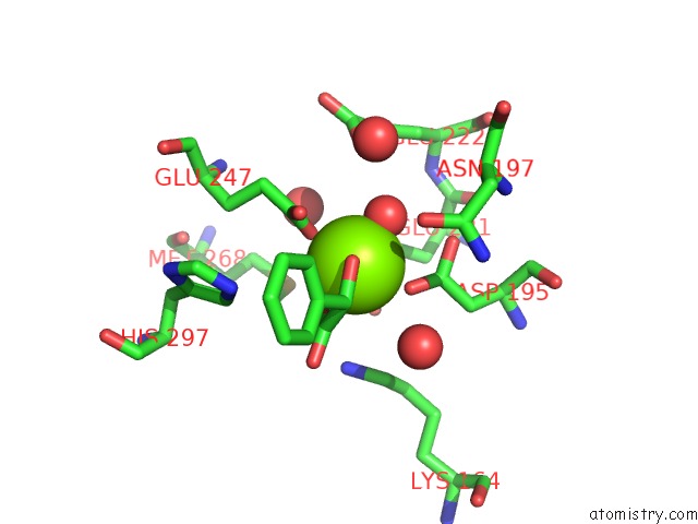
Mono view
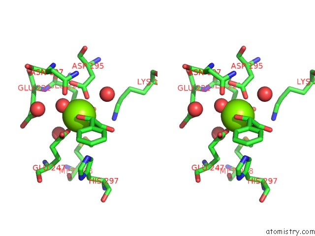
Stereo pair view
Mono view
Stereo pair view
A full contact list of Magnesium with other atoms in the Mg binding
site number 1 of Mandelate Racemase Mutant K166R Co-Crystallized with (R)-Mandelate within 5.0Å range:
|
Reference:
A.T.Kallarakal,
B.Mitra,
J.W.Kozarich,
J.A.Gerlt,
J.G.Clifton,
G.A.Petsko,
G.L.Kenyon.
Mechanism of the Reaction Catalyzed By Mandelate Racemase: Structure and Mechanistic Properties of the K166R Mutant. Biochemistry V. 34 2788 1995.
ISSN: ISSN 0006-2960
PubMed: 7893690
DOI: 10.1021/BI00009A007
Page generated: Sun Aug 10 00:50:17 2025
ISSN: ISSN 0006-2960
PubMed: 7893690
DOI: 10.1021/BI00009A007
Last articles
Mg in 3J7HMg in 3J7I
Mg in 3IVK
Mg in 3J6E
Mg in 3J6G
Mg in 3J6P
Mg in 3J6H
Mg in 3J1F
Mg in 3J6F
Mg in 3J5V