Magnesium »
PDB 1mum-1n6i »
1n5k »
Magnesium in PDB 1n5k: Crystal Structure of Mycobacterium Tuberculosis Thymidylate Kinase Crystallized in Sodium Malonate (Resolution 2.1 A)
Enzymatic activity of Crystal Structure of Mycobacterium Tuberculosis Thymidylate Kinase Crystallized in Sodium Malonate (Resolution 2.1 A)
All present enzymatic activity of Crystal Structure of Mycobacterium Tuberculosis Thymidylate Kinase Crystallized in Sodium Malonate (Resolution 2.1 A):
2.7.4.9;
2.7.4.9;
Protein crystallography data
The structure of Crystal Structure of Mycobacterium Tuberculosis Thymidylate Kinase Crystallized in Sodium Malonate (Resolution 2.1 A), PDB code: 1n5k
was solved by
E.Fioravanti,
A.Haouz,
T.Ursby,
H.Munier-Lehmann,
M.Delarue,
D.Bourgeois,
with X-Ray Crystallography technique. A brief refinement statistics is given in the table below:
| Resolution Low / High (Å) | 19.71 / 2.10 |
| Space group | P 31 2 1 |
| Cell size a, b, c (Å), α, β, γ (°) | 64.254, 64.254, 195.484, 90.00, 90.00, 120.00 |
| R / Rfree (%) | 21.5 / 24.3 |
Magnesium Binding Sites:
The binding sites of Magnesium atom in the Crystal Structure of Mycobacterium Tuberculosis Thymidylate Kinase Crystallized in Sodium Malonate (Resolution 2.1 A)
(pdb code 1n5k). This binding sites where shown within
5.0 Angstroms radius around Magnesium atom.
In total only one binding site of Magnesium was determined in the Crystal Structure of Mycobacterium Tuberculosis Thymidylate Kinase Crystallized in Sodium Malonate (Resolution 2.1 A), PDB code: 1n5k:
In total only one binding site of Magnesium was determined in the Crystal Structure of Mycobacterium Tuberculosis Thymidylate Kinase Crystallized in Sodium Malonate (Resolution 2.1 A), PDB code: 1n5k:
Magnesium binding site 1 out of 1 in 1n5k
Go back to
Magnesium binding site 1 out
of 1 in the Crystal Structure of Mycobacterium Tuberculosis Thymidylate Kinase Crystallized in Sodium Malonate (Resolution 2.1 A)
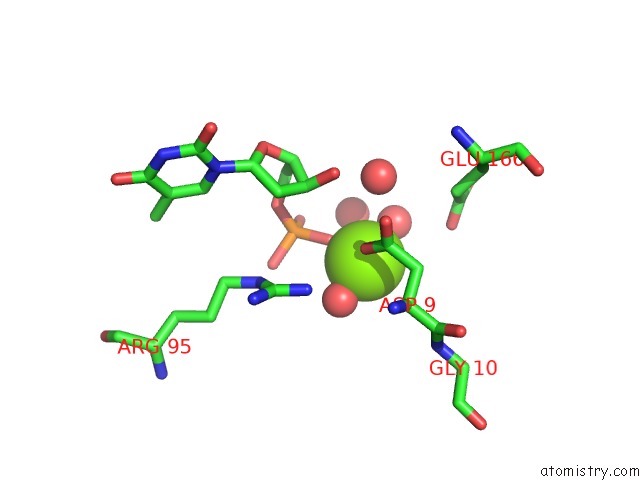
Mono view
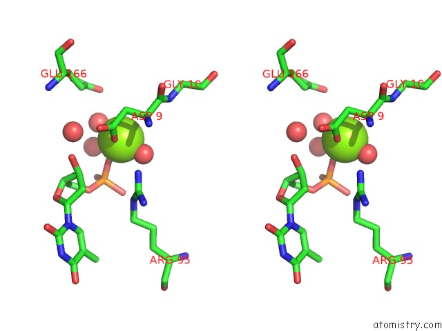
Stereo pair view
Mono view
Stereo pair view
A full contact list of Magnesium with other atoms in the Mg binding
site number 1 of Crystal Structure of Mycobacterium Tuberculosis Thymidylate Kinase Crystallized in Sodium Malonate (Resolution 2.1 A) within 5.0Å range:
|
Reference:
E.Fioravanti,
A.Haouz,
T.Ursby,
H.Munier-Lehmann,
M.Delarue,
D.Bourgeois.
Mycobacterium Tuberculosis Thymidylate Kinase: Structural Studies of Intermediates Along the Reaction Pathway J.Mol.Biol. V. 375 1077 2003.
ISSN: ISSN 0022-2836
PubMed: 12662932
DOI: 10.1016/S0022-2836(03)00202-X
Page generated: Sun Aug 10 01:18:49 2025
ISSN: ISSN 0022-2836
PubMed: 12662932
DOI: 10.1016/S0022-2836(03)00202-X
Last articles
Mg in 2OQYMg in 2OUP
Mg in 2OUN
Mg in 2OU7
Mg in 2OTG
Mg in 2OSB
Mg in 2OS8
Mg in 2ORI
Mg in 2ORW
Mg in 2ONP