Magnesium »
PDB 1qc5-1qsh »
1qs0 »
Magnesium in PDB 1qs0: Crystal Structure of Pseudomonas Putida 2-Oxoisovalerate Dehydrogenase (Branched-Chain Alpha-Keto Acid Dehydrogenase, E1B)
Protein crystallography data
The structure of Crystal Structure of Pseudomonas Putida 2-Oxoisovalerate Dehydrogenase (Branched-Chain Alpha-Keto Acid Dehydrogenase, E1B), PDB code: 1qs0
was solved by
A.Aevarsson,
K.Seger,
S.Turley,
J.R.Sokatch,
W.G.J.Hol,
with X-Ray Crystallography technique. A brief refinement statistics is given in the table below:
| Resolution Low / High (Å) | 40.00 / 2.40 |
| Space group | I 41 2 2 |
| Cell size a, b, c (Å), α, β, γ (°) | 101.340, 101.340, 381.230, 90.00, 90.00, 90.00 |
| R / Rfree (%) | 21.8 / 26.5 |
Magnesium Binding Sites:
The binding sites of Magnesium atom in the Crystal Structure of Pseudomonas Putida 2-Oxoisovalerate Dehydrogenase (Branched-Chain Alpha-Keto Acid Dehydrogenase, E1B)
(pdb code 1qs0). This binding sites where shown within
5.0 Angstroms radius around Magnesium atom.
In total only one binding site of Magnesium was determined in the Crystal Structure of Pseudomonas Putida 2-Oxoisovalerate Dehydrogenase (Branched-Chain Alpha-Keto Acid Dehydrogenase, E1B), PDB code: 1qs0:
In total only one binding site of Magnesium was determined in the Crystal Structure of Pseudomonas Putida 2-Oxoisovalerate Dehydrogenase (Branched-Chain Alpha-Keto Acid Dehydrogenase, E1B), PDB code: 1qs0:
Magnesium binding site 1 out of 1 in 1qs0
Go back to
Magnesium binding site 1 out
of 1 in the Crystal Structure of Pseudomonas Putida 2-Oxoisovalerate Dehydrogenase (Branched-Chain Alpha-Keto Acid Dehydrogenase, E1B)
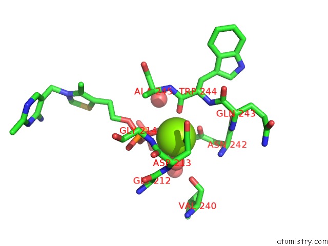
Mono view
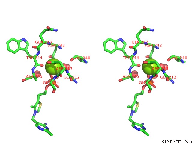
Stereo pair view
Mono view
Stereo pair view
A full contact list of Magnesium with other atoms in the Mg binding
site number 1 of Crystal Structure of Pseudomonas Putida 2-Oxoisovalerate Dehydrogenase (Branched-Chain Alpha-Keto Acid Dehydrogenase, E1B) within 5.0Å range:
|
Reference:
A.Aevarsson,
K.Seger,
S.Turley,
J.R.Sokatch,
W.G.Hol.
Crystal Structure of 2-Oxoisovalerate and Dehydrogenase and the Architecture of 2-Oxo Acid Dehydrogenase Multienzyme Complexes. Nat.Struct.Biol. V. 6 785 1999.
ISSN: ISSN 1072-8368
PubMed: 10426958
DOI: 10.1038/11563
Page generated: Sun Aug 10 03:05:35 2025
ISSN: ISSN 1072-8368
PubMed: 10426958
DOI: 10.1038/11563
Last articles
Mg in 1VQ7Mg in 1VQ5
Mg in 1VQ6
Mg in 1VQ8
Mg in 1VQ4
Mg in 1VPA
Mg in 1VPE
Mg in 1VOM
Mg in 1VMA
Mg in 1VMK