Magnesium »
PDB 1so6-1t5s »
1sww »
Magnesium in PDB 1sww: Crystal Structure of the Phosphonoacetaldehyde Hydrolase D12A Mutant Complexed with Magnesium and Substrate Phosphonoacetaldehyde
Enzymatic activity of Crystal Structure of the Phosphonoacetaldehyde Hydrolase D12A Mutant Complexed with Magnesium and Substrate Phosphonoacetaldehyde
All present enzymatic activity of Crystal Structure of the Phosphonoacetaldehyde Hydrolase D12A Mutant Complexed with Magnesium and Substrate Phosphonoacetaldehyde:
3.11.1.1;
3.11.1.1;
Protein crystallography data
The structure of Crystal Structure of the Phosphonoacetaldehyde Hydrolase D12A Mutant Complexed with Magnesium and Substrate Phosphonoacetaldehyde, PDB code: 1sww
was solved by
G.Zhang,
M.C.Morais,
J.Dai,
W.Zhang,
D.Dunaway-Mariano,
K.N.Allen,
with X-Ray Crystallography technique. A brief refinement statistics is given in the table below:
| Resolution Low / High (Å) | 60.68 / 2.30 |
| Space group | P 1 21 1 |
| Cell size a, b, c (Å), α, β, γ (°) | 63.815, 62.520, 72.618, 90.00, 108.03, 90.00 |
| R / Rfree (%) | 21.8 / 27.8 |
Magnesium Binding Sites:
The binding sites of Magnesium atom in the Crystal Structure of the Phosphonoacetaldehyde Hydrolase D12A Mutant Complexed with Magnesium and Substrate Phosphonoacetaldehyde
(pdb code 1sww). This binding sites where shown within
5.0 Angstroms radius around Magnesium atom.
In total 2 binding sites of Magnesium where determined in the Crystal Structure of the Phosphonoacetaldehyde Hydrolase D12A Mutant Complexed with Magnesium and Substrate Phosphonoacetaldehyde, PDB code: 1sww:
Jump to Magnesium binding site number: 1; 2;
In total 2 binding sites of Magnesium where determined in the Crystal Structure of the Phosphonoacetaldehyde Hydrolase D12A Mutant Complexed with Magnesium and Substrate Phosphonoacetaldehyde, PDB code: 1sww:
Jump to Magnesium binding site number: 1; 2;
Magnesium binding site 1 out of 2 in 1sww
Go back to
Magnesium binding site 1 out
of 2 in the Crystal Structure of the Phosphonoacetaldehyde Hydrolase D12A Mutant Complexed with Magnesium and Substrate Phosphonoacetaldehyde
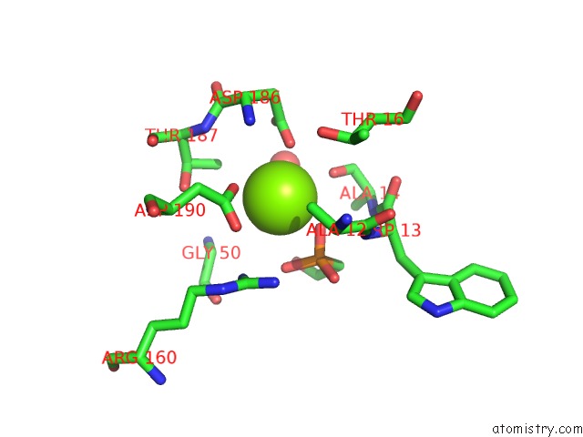
Mono view
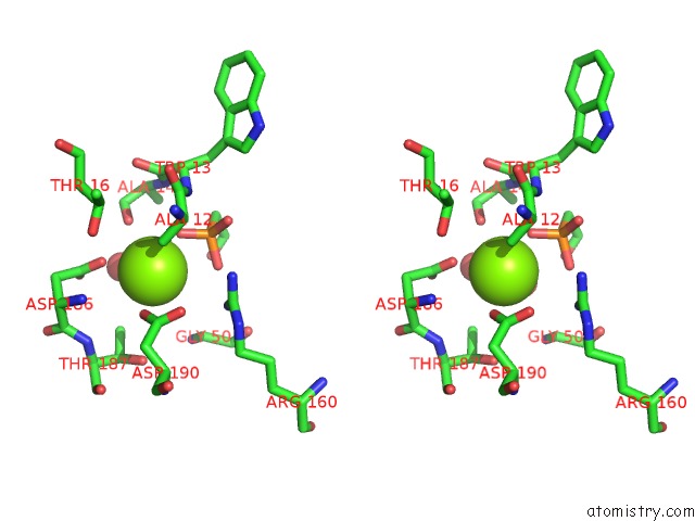
Stereo pair view
Mono view
Stereo pair view
A full contact list of Magnesium with other atoms in the Mg binding
site number 1 of Crystal Structure of the Phosphonoacetaldehyde Hydrolase D12A Mutant Complexed with Magnesium and Substrate Phosphonoacetaldehyde within 5.0Å range:
|
Magnesium binding site 2 out of 2 in 1sww
Go back to
Magnesium binding site 2 out
of 2 in the Crystal Structure of the Phosphonoacetaldehyde Hydrolase D12A Mutant Complexed with Magnesium and Substrate Phosphonoacetaldehyde
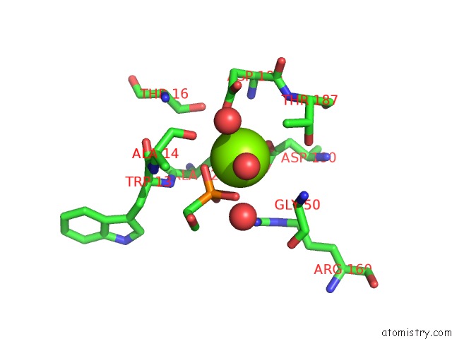
Mono view
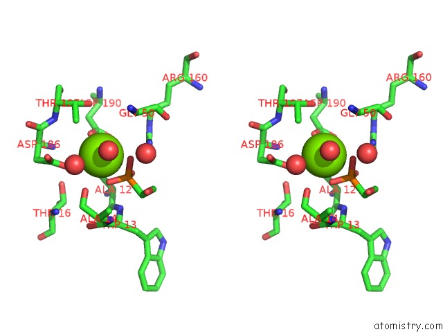
Stereo pair view
Mono view
Stereo pair view
A full contact list of Magnesium with other atoms in the Mg binding
site number 2 of Crystal Structure of the Phosphonoacetaldehyde Hydrolase D12A Mutant Complexed with Magnesium and Substrate Phosphonoacetaldehyde within 5.0Å range:
|
Reference:
G.Zhang,
M.C.Morais,
J.Dai,
W.Zhang,
D.Dunaway-Mariano,
K.N.Allen.
Investigation of Metal Ion Binding in Phosphonoacetaldehyde Hydrolase Identifies Sequence Markers For Metal-Activated Enzymes of the Had Enzyme Superfamily Biochemistry V. 43 4990 2004.
ISSN: ISSN 0006-2960
PubMed: 15109258
DOI: 10.1021/BI036309N
Page generated: Sun Aug 10 04:46:00 2025
ISSN: ISSN 0006-2960
PubMed: 15109258
DOI: 10.1021/BI036309N
Last articles
Mg in 2CW0Mg in 2CZ1
Mg in 2CYZ
Mg in 2CVY
Mg in 2CVX
Mg in 2CVW
Mg in 2CVV
Mg in 2CVU
Mg in 2CV1
Mg in 2CVT