Magnesium »
PDB 1ueu-1v5f »
1un9 »
Magnesium in PDB 1un9: Crystal Structure of the Dihydroxyacetone Kinase From C. Freundii in Complex with Amp-Pnp and MG2+
Enzymatic activity of Crystal Structure of the Dihydroxyacetone Kinase From C. Freundii in Complex with Amp-Pnp and MG2+
All present enzymatic activity of Crystal Structure of the Dihydroxyacetone Kinase From C. Freundii in Complex with Amp-Pnp and MG2+:
2.7.1.29;
2.7.1.29;
Protein crystallography data
The structure of Crystal Structure of the Dihydroxyacetone Kinase From C. Freundii in Complex with Amp-Pnp and MG2+, PDB code: 1un9
was solved by
C.Siebold,
I.Arnold,
L.F.Garcia-Alles,
U.Baumann,
B.Erni,
with X-Ray Crystallography technique. A brief refinement statistics is given in the table below:
| Resolution Low / High (Å) | 25 / 3.1 |
| Space group | C 2 2 21 |
| Cell size a, b, c (Å), α, β, γ (°) | 100.788, 124.869, 236.530, 90.00, 90.00, 90.00 |
| R / Rfree (%) | 23.7 / n/a |
Magnesium Binding Sites:
The binding sites of Magnesium atom in the Crystal Structure of the Dihydroxyacetone Kinase From C. Freundii in Complex with Amp-Pnp and MG2+
(pdb code 1un9). This binding sites where shown within
5.0 Angstroms radius around Magnesium atom.
In total 4 binding sites of Magnesium where determined in the Crystal Structure of the Dihydroxyacetone Kinase From C. Freundii in Complex with Amp-Pnp and MG2+, PDB code: 1un9:
Jump to Magnesium binding site number: 1; 2; 3; 4;
In total 4 binding sites of Magnesium where determined in the Crystal Structure of the Dihydroxyacetone Kinase From C. Freundii in Complex with Amp-Pnp and MG2+, PDB code: 1un9:
Jump to Magnesium binding site number: 1; 2; 3; 4;
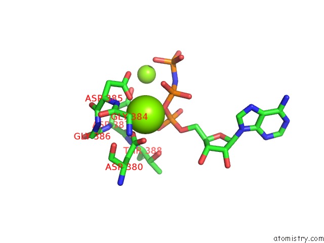
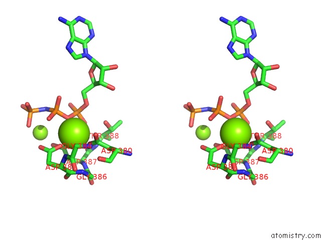
Magnesium binding site 1 out of 4 in 1un9
Go back to
Magnesium binding site 1 out
of 4 in the Crystal Structure of the Dihydroxyacetone Kinase From C. Freundii in Complex with Amp-Pnp and MG2+
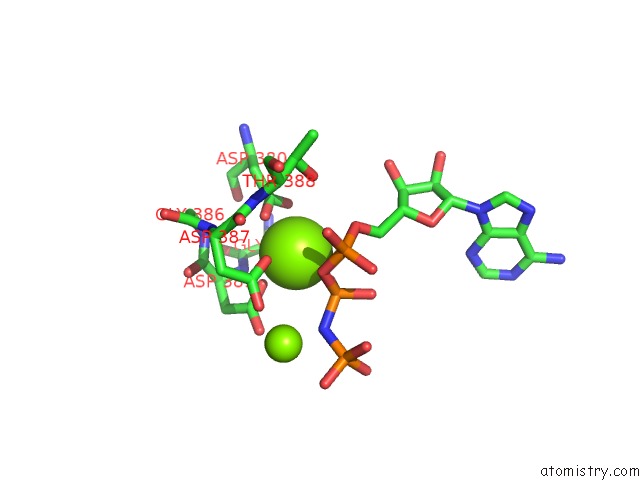
Mono view
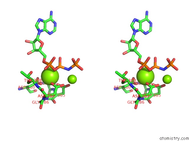
Stereo pair view
Mono view
Stereo pair view
A full contact list of Magnesium with other atoms in the Mg binding
site number 1 of Crystal Structure of the Dihydroxyacetone Kinase From C. Freundii in Complex with Amp-Pnp and MG2+ within 5.0Å range:
|
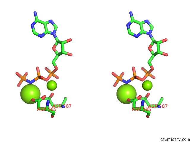
Magnesium binding site 2 out of 4 in 1un9
Go back to
Magnesium binding site 2 out
of 4 in the Crystal Structure of the Dihydroxyacetone Kinase From C. Freundii in Complex with Amp-Pnp and MG2+
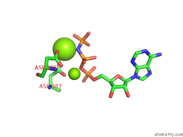
Mono view
Stereo pair view
Mono view
Stereo pair view
A full contact list of Magnesium with other atoms in the Mg binding
site number 2 of Crystal Structure of the Dihydroxyacetone Kinase From C. Freundii in Complex with Amp-Pnp and MG2+ within 5.0Å range:
|
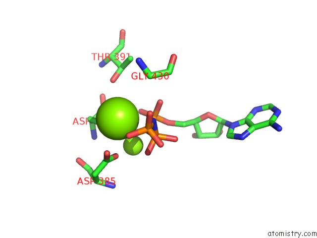
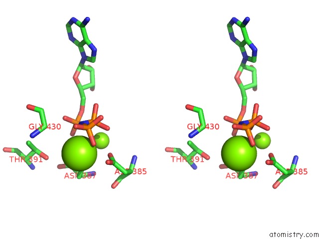
Magnesium binding site 3 out of 4 in 1un9
Go back to
Magnesium binding site 3 out
of 4 in the Crystal Structure of the Dihydroxyacetone Kinase From C. Freundii in Complex with Amp-Pnp and MG2+
Mono view
Stereo pair view
Mono view
Stereo pair view
A full contact list of Magnesium with other atoms in the Mg binding
site number 3 of Crystal Structure of the Dihydroxyacetone Kinase From C. Freundii in Complex with Amp-Pnp and MG2+ within 5.0Å range:
|
Magnesium binding site 4 out of 4 in 1un9
Go back to
Magnesium binding site 4 out
of 4 in the Crystal Structure of the Dihydroxyacetone Kinase From C. Freundii in Complex with Amp-Pnp and MG2+
Mono view
Stereo pair view
Mono view
Stereo pair view
A full contact list of Magnesium with other atoms in the Mg binding
site number 4 of Crystal Structure of the Dihydroxyacetone Kinase From C. Freundii in Complex with Amp-Pnp and MG2+ within 5.0Å range:
|
Reference:
C.Siebold,
I.Arnold,
L.F.Garcia-Alles,
U.Baumann,
B.Erni.
Crystal Structure of the Citrobacter Freundii Dihydroxyacetone Kinase Reveals An Eight-Stranded Alpha-Helical Barrel Atp-Binding Domain J.Biol.Chem. V. 278 48236 2003.
ISSN: ISSN 0021-9258
PubMed: 12966101
DOI: 10.1074/JBC.M305942200
Page generated: Tue Aug 13 14:52:40 2024
ISSN: ISSN 0021-9258
PubMed: 12966101
DOI: 10.1074/JBC.M305942200
Last articles
Zn in 9J0NZn in 9J0O
Zn in 9J0P
Zn in 9FJX
Zn in 9EKB
Zn in 9C0F
Zn in 9CAH
Zn in 9CH0
Zn in 9CH3
Zn in 9CH1