Magnesium »
PDB 1x1t-1xfx »
1xed »
Magnesium in PDB 1xed: Crystal Structure of A Ligand-Binding Domain of the Human Polymeric Ig Receptor, Pigr
Protein crystallography data
The structure of Crystal Structure of A Ligand-Binding Domain of the Human Polymeric Ig Receptor, Pigr, PDB code: 1xed
was solved by
A.E.Hamburger,
A.P.West Jr.,
P.J.Bjorkman,
with X-Ray Crystallography technique. A brief refinement statistics is given in the table below:
| Resolution Low / High (Å) | 20.00 / 1.90 |
| Space group | P 1 21 1 |
| Cell size a, b, c (Å), α, β, γ (°) | 41.996, 157.195, 53.860, 90.00, 112.94, 90.00 |
| R / Rfree (%) | 18.3 / 24.4 |
Magnesium Binding Sites:
The binding sites of Magnesium atom in the Crystal Structure of A Ligand-Binding Domain of the Human Polymeric Ig Receptor, Pigr
(pdb code 1xed). This binding sites where shown within
5.0 Angstroms radius around Magnesium atom.
In total 2 binding sites of Magnesium where determined in the Crystal Structure of A Ligand-Binding Domain of the Human Polymeric Ig Receptor, Pigr, PDB code: 1xed:
Jump to Magnesium binding site number: 1; 2;
In total 2 binding sites of Magnesium where determined in the Crystal Structure of A Ligand-Binding Domain of the Human Polymeric Ig Receptor, Pigr, PDB code: 1xed:
Jump to Magnesium binding site number: 1; 2;
Magnesium binding site 1 out of 2 in 1xed
Go back to
Magnesium binding site 1 out
of 2 in the Crystal Structure of A Ligand-Binding Domain of the Human Polymeric Ig Receptor, Pigr
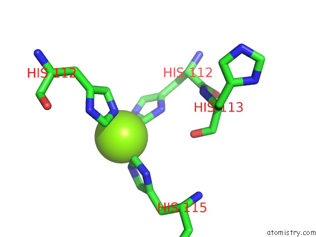
Mono view
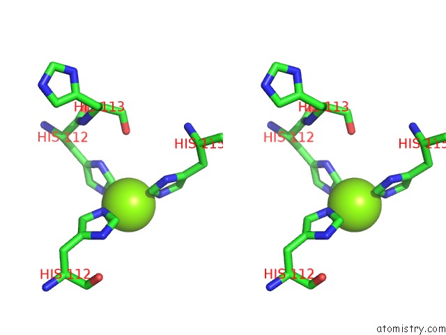
Stereo pair view
Mono view
Stereo pair view
A full contact list of Magnesium with other atoms in the Mg binding
site number 1 of Crystal Structure of A Ligand-Binding Domain of the Human Polymeric Ig Receptor, Pigr within 5.0Å range:
|
Magnesium binding site 2 out of 2 in 1xed
Go back to
Magnesium binding site 2 out
of 2 in the Crystal Structure of A Ligand-Binding Domain of the Human Polymeric Ig Receptor, Pigr
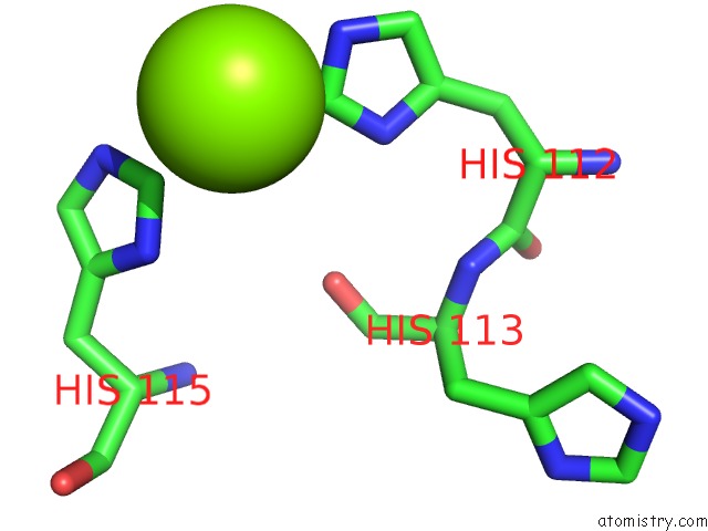
Mono view
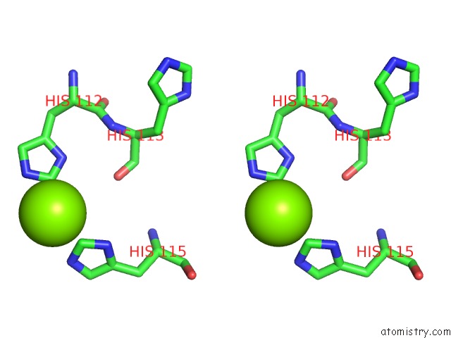
Stereo pair view
Mono view
Stereo pair view
A full contact list of Magnesium with other atoms in the Mg binding
site number 2 of Crystal Structure of A Ligand-Binding Domain of the Human Polymeric Ig Receptor, Pigr within 5.0Å range:
|
Reference:
A.E.Hamburger,
A.P.West Jr.,
P.J.Bjorkman.
Crystal Structure of A Polymeric Immunoglobulin Binding Fragment of the Human Polymeric Immunoglobulin Receptor Structure V. 12 1925 2004.
ISSN: ISSN 0969-2126
PubMed: 15530357
DOI: 10.1016/J.STR.2004.09.006
Page generated: Tue Aug 13 17:32:40 2024
ISSN: ISSN 0969-2126
PubMed: 15530357
DOI: 10.1016/J.STR.2004.09.006
Last articles
Zn in 9J0NZn in 9J0O
Zn in 9J0P
Zn in 9FJX
Zn in 9EKB
Zn in 9C0F
Zn in 9CAH
Zn in 9CH0
Zn in 9CH3
Zn in 9CH1