Magnesium »
PDB 1xyl-1ydf »
1y0q »
Magnesium in PDB 1y0q: Crystal Structure of An Active Group I Ribozyme-Product Complex
Protein crystallography data
The structure of Crystal Structure of An Active Group I Ribozyme-Product Complex, PDB code: 1y0q
was solved by
B.L.Golden,
H.Kim,
E.Chase,
with X-Ray Crystallography technique. A brief refinement statistics is given in the table below:
| Resolution Low / High (Å) | 19.54 / 3.60 |
| Space group | I 21 21 21 |
| Cell size a, b, c (Å), α, β, γ (°) | 94.570, 140.970, 210.850, 90.00, 90.00, 90.00 |
| R / Rfree (%) | 27.7 / 31 |
Magnesium Binding Sites:
The binding sites of Magnesium atom in the Crystal Structure of An Active Group I Ribozyme-Product Complex
(pdb code 1y0q). This binding sites where shown within
5.0 Angstroms radius around Magnesium atom.
In total 4 binding sites of Magnesium where determined in the Crystal Structure of An Active Group I Ribozyme-Product Complex, PDB code: 1y0q:
Jump to Magnesium binding site number: 1; 2; 3; 4;
In total 4 binding sites of Magnesium where determined in the Crystal Structure of An Active Group I Ribozyme-Product Complex, PDB code: 1y0q:
Jump to Magnesium binding site number: 1; 2; 3; 4;
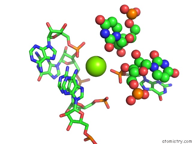
Magnesium binding site 1 out of 4 in 1y0q
Go back to
Magnesium binding site 1 out
of 4 in the Crystal Structure of An Active Group I Ribozyme-Product Complex
Mono view

Stereo pair view
Mono view
Stereo pair view
A full contact list of Magnesium with other atoms in the Mg binding
site number 1 of Crystal Structure of An Active Group I Ribozyme-Product Complex within 5.0Å range:
|
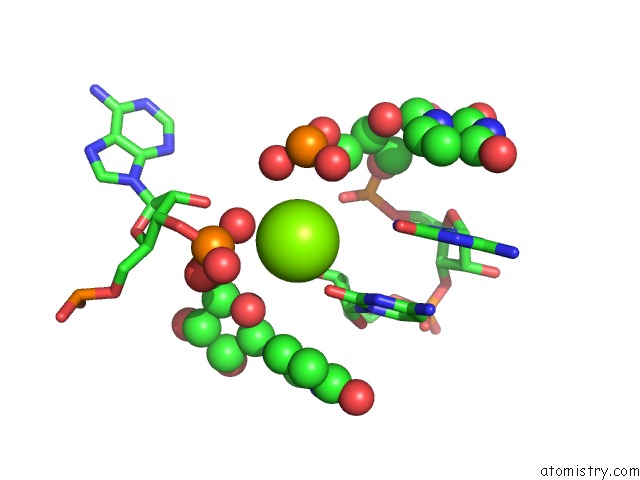
Magnesium binding site 2 out of 4 in 1y0q
Go back to
Magnesium binding site 2 out
of 4 in the Crystal Structure of An Active Group I Ribozyme-Product Complex
Mono view
Stereo pair view
Mono view
Stereo pair view
A full contact list of Magnesium with other atoms in the Mg binding
site number 2 of Crystal Structure of An Active Group I Ribozyme-Product Complex within 5.0Å range:
|
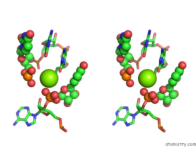
Magnesium binding site 3 out of 4 in 1y0q
Go back to
Magnesium binding site 3 out
of 4 in the Crystal Structure of An Active Group I Ribozyme-Product Complex
Mono view

Stereo pair view
Mono view
Stereo pair view
A full contact list of Magnesium with other atoms in the Mg binding
site number 3 of Crystal Structure of An Active Group I Ribozyme-Product Complex within 5.0Å range:
|
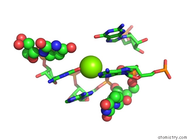
Magnesium binding site 4 out of 4 in 1y0q
Go back to
Magnesium binding site 4 out
of 4 in the Crystal Structure of An Active Group I Ribozyme-Product Complex

Mono view

Stereo pair view
Mono view
Stereo pair view
A full contact list of Magnesium with other atoms in the Mg binding
site number 4 of Crystal Structure of An Active Group I Ribozyme-Product Complex within 5.0Å range:
|
Reference:
B.L.Golden,
H.Kim,
E.Chase.
Crystal Structure of A Phage Twort Group I Ribozyme-Product Complex Nat.Struct.Mol.Biol. V. 12 82 2005.
ISSN: ISSN 1545-9993
PubMed: 15580277
DOI: 10.1038/NSMB868
Page generated: Tue Aug 13 18:27:58 2024
ISSN: ISSN 1545-9993
PubMed: 15580277
DOI: 10.1038/NSMB868
Last articles
Zn in 9MJ5Zn in 9HNW
Zn in 9G0L
Zn in 9FNE
Zn in 9DZN
Zn in 9E0I
Zn in 9D32
Zn in 9DAK
Zn in 8ZXC
Zn in 8ZUF