Magnesium »
PDB 2hld-2hxf »
2hvq »
Magnesium in PDB 2hvq: Structure of Adenylated Full-Length T4 Rna Ligase 2
Protein crystallography data
The structure of Structure of Adenylated Full-Length T4 Rna Ligase 2, PDB code: 2hvq
was solved by
J.Nandakumar,
C.D.Lima,
with X-Ray Crystallography technique. A brief refinement statistics is given in the table below:
| Resolution Low / High (Å) | 41.56 / 2.40 |
| Space group | P 21 21 21 |
| Cell size a, b, c (Å), α, β, γ (°) | 48.020, 57.850, 119.490, 90.00, 90.00, 90.00 |
| R / Rfree (%) | 21.1 / 27.4 |
Magnesium Binding Sites:
The binding sites of Magnesium atom in the Structure of Adenylated Full-Length T4 Rna Ligase 2
(pdb code 2hvq). This binding sites where shown within
5.0 Angstroms radius around Magnesium atom.
In total 2 binding sites of Magnesium where determined in the Structure of Adenylated Full-Length T4 Rna Ligase 2, PDB code: 2hvq:
Jump to Magnesium binding site number: 1; 2;
In total 2 binding sites of Magnesium where determined in the Structure of Adenylated Full-Length T4 Rna Ligase 2, PDB code: 2hvq:
Jump to Magnesium binding site number: 1; 2;
Magnesium binding site 1 out of 2 in 2hvq
Go back to
Magnesium binding site 1 out
of 2 in the Structure of Adenylated Full-Length T4 Rna Ligase 2
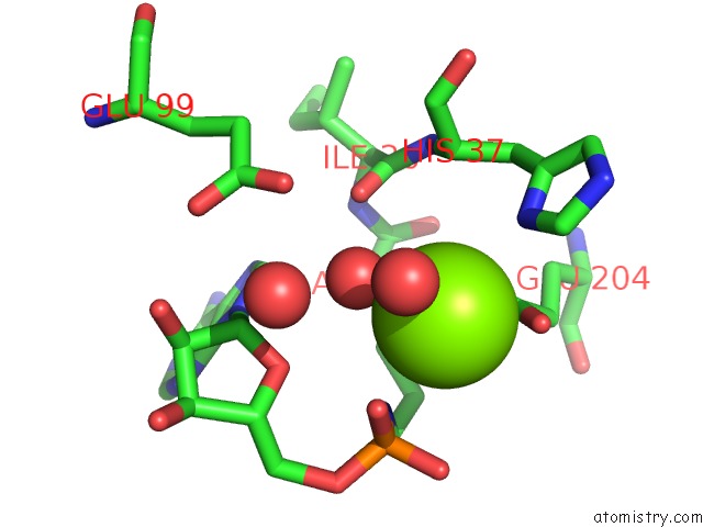
Mono view
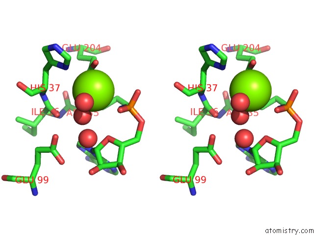
Stereo pair view
Mono view
Stereo pair view
A full contact list of Magnesium with other atoms in the Mg binding
site number 1 of Structure of Adenylated Full-Length T4 Rna Ligase 2 within 5.0Å range:
|
Magnesium binding site 2 out of 2 in 2hvq
Go back to
Magnesium binding site 2 out
of 2 in the Structure of Adenylated Full-Length T4 Rna Ligase 2
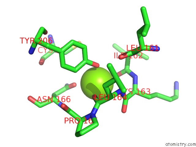
Mono view
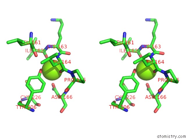
Stereo pair view
Mono view
Stereo pair view
A full contact list of Magnesium with other atoms in the Mg binding
site number 2 of Structure of Adenylated Full-Length T4 Rna Ligase 2 within 5.0Å range:
|
Reference:
J.Nandakumar,
S.Shuman,
C.D.Lima.
Rna Ligase Structures Reveal the Basis For Rna Specificity and Conformational Changes That Drive Ligation Forward. Cell(Cambridge,Mass.) V. 127 71 2006.
ISSN: ISSN 0092-8674
PubMed: 17018278
DOI: 10.1016/J.CELL.2006.08.038
Page generated: Sun Aug 10 11:28:15 2025
ISSN: ISSN 0092-8674
PubMed: 17018278
DOI: 10.1016/J.CELL.2006.08.038
Last articles
Mg in 6EVWMg in 6EVY
Mg in 6EVX
Mg in 6EU3
Mg in 6EU2
Mg in 6EPZ
Mg in 6EU1
Mg in 6EU0
Mg in 6ETH
Mg in 6ETF