Magnesium »
PDB 2hld-2hxf »
2hwg »
Magnesium in PDB 2hwg: Structure of Phosphorylated Enzyme I of the Phosphoenolpyruvate:Sugar Phosphotransferase System
Enzymatic activity of Structure of Phosphorylated Enzyme I of the Phosphoenolpyruvate:Sugar Phosphotransferase System
All present enzymatic activity of Structure of Phosphorylated Enzyme I of the Phosphoenolpyruvate:Sugar Phosphotransferase System:
2.7.3.9;
2.7.3.9;
Protein crystallography data
The structure of Structure of Phosphorylated Enzyme I of the Phosphoenolpyruvate:Sugar Phosphotransferase System, PDB code: 2hwg
was solved by
K.Lim,
A.Teplyakov,
O.Herzberg,
with X-Ray Crystallography technique. A brief refinement statistics is given in the table below:
| Resolution Low / High (Å) | 50.00 / 2.70 |
| Space group | P 21 21 21 |
| Cell size a, b, c (Å), α, β, γ (°) | 85.487, 94.084, 161.007, 90.00, 90.00, 90.00 |
| R / Rfree (%) | 20.4 / 28.4 |
Magnesium Binding Sites:
The binding sites of Magnesium atom in the Structure of Phosphorylated Enzyme I of the Phosphoenolpyruvate:Sugar Phosphotransferase System
(pdb code 2hwg). This binding sites where shown within
5.0 Angstroms radius around Magnesium atom.
In total 2 binding sites of Magnesium where determined in the Structure of Phosphorylated Enzyme I of the Phosphoenolpyruvate:Sugar Phosphotransferase System, PDB code: 2hwg:
Jump to Magnesium binding site number: 1; 2;
In total 2 binding sites of Magnesium where determined in the Structure of Phosphorylated Enzyme I of the Phosphoenolpyruvate:Sugar Phosphotransferase System, PDB code: 2hwg:
Jump to Magnesium binding site number: 1; 2;
Magnesium binding site 1 out of 2 in 2hwg
Go back to
Magnesium binding site 1 out
of 2 in the Structure of Phosphorylated Enzyme I of the Phosphoenolpyruvate:Sugar Phosphotransferase System
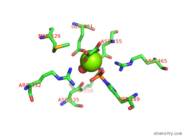
Mono view
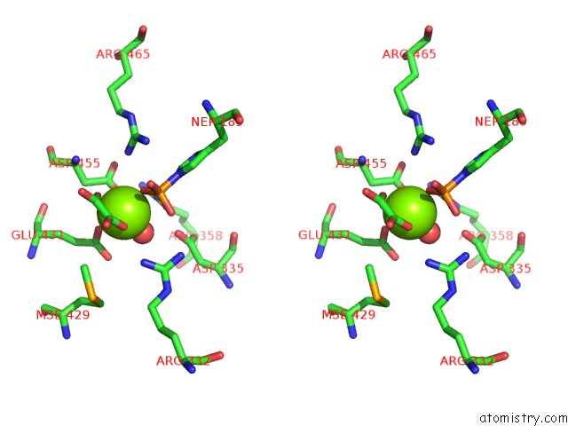
Stereo pair view
Mono view
Stereo pair view
A full contact list of Magnesium with other atoms in the Mg binding
site number 1 of Structure of Phosphorylated Enzyme I of the Phosphoenolpyruvate:Sugar Phosphotransferase System within 5.0Å range:
|
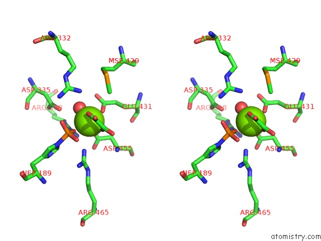
Magnesium binding site 2 out of 2 in 2hwg
Go back to
Magnesium binding site 2 out
of 2 in the Structure of Phosphorylated Enzyme I of the Phosphoenolpyruvate:Sugar Phosphotransferase System
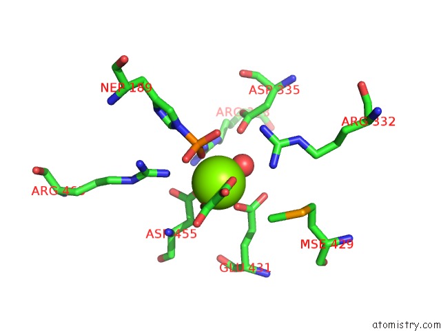
Mono view
Stereo pair view
Mono view
Stereo pair view
A full contact list of Magnesium with other atoms in the Mg binding
site number 2 of Structure of Phosphorylated Enzyme I of the Phosphoenolpyruvate:Sugar Phosphotransferase System within 5.0Å range:
|
Reference:
A.Teplyakov,
K.Lim,
P.P.Zhu,
G.Kapadia,
C.C.Chen,
J.Schwartz,
A.Howard,
P.T.Reddy,
A.Peterkofsky,
O.Herzberg.
Structure of Phosphorylated Enzyme I, the Phosphoenolpyruvate:Sugar Phosphotransferase System Sugar Translocation Signal Protein. Proc.Natl.Acad.Sci.Usa V. 103 16218 2006.
ISSN: ISSN 0027-8424
PubMed: 17053069
DOI: 10.1073/PNAS.0607587103
Page generated: Sun Aug 10 11:29:05 2025
ISSN: ISSN 0027-8424
PubMed: 17053069
DOI: 10.1073/PNAS.0607587103
Last articles
Mg in 5XTMMg in 5XUT
Mg in 5XUS
Mg in 5XUJ
Mg in 5XUI
Mg in 5XU1
Mg in 5XT8
Mg in 5XT2
Mg in 5XR7
Mg in 5XR6