Magnesium »
PDB 2o56-2oh6 »
2o8f »
Magnesium in PDB 2o8f: Human Mutsalpha (MSH2/MSH6) Bound to Dna with A Single Base T Insert
Protein crystallography data
The structure of Human Mutsalpha (MSH2/MSH6) Bound to Dna with A Single Base T Insert, PDB code: 2o8f
was solved by
J.J.Warren,
T.J.Pohlhaus,
A.Changela,
P.L.Modrich,
L.S.Beese,
with X-Ray Crystallography technique. A brief refinement statistics is given in the table below:
| Resolution Low / High (Å) | 20.00 / 3.25 |
| Space group | P 43 3 2 |
| Cell size a, b, c (Å), α, β, γ (°) | 259.550, 259.550, 259.550, 90.00, 90.00, 90.00 |
| R / Rfree (%) | 24.3 / 29.3 |
Magnesium Binding Sites:
The binding sites of Magnesium atom in the Human Mutsalpha (MSH2/MSH6) Bound to Dna with A Single Base T Insert
(pdb code 2o8f). This binding sites where shown within
5.0 Angstroms radius around Magnesium atom.
In total only one binding site of Magnesium was determined in the Human Mutsalpha (MSH2/MSH6) Bound to Dna with A Single Base T Insert, PDB code: 2o8f:
In total only one binding site of Magnesium was determined in the Human Mutsalpha (MSH2/MSH6) Bound to Dna with A Single Base T Insert, PDB code: 2o8f:
Magnesium binding site 1 out of 1 in 2o8f
Go back to
Magnesium binding site 1 out
of 1 in the Human Mutsalpha (MSH2/MSH6) Bound to Dna with A Single Base T Insert
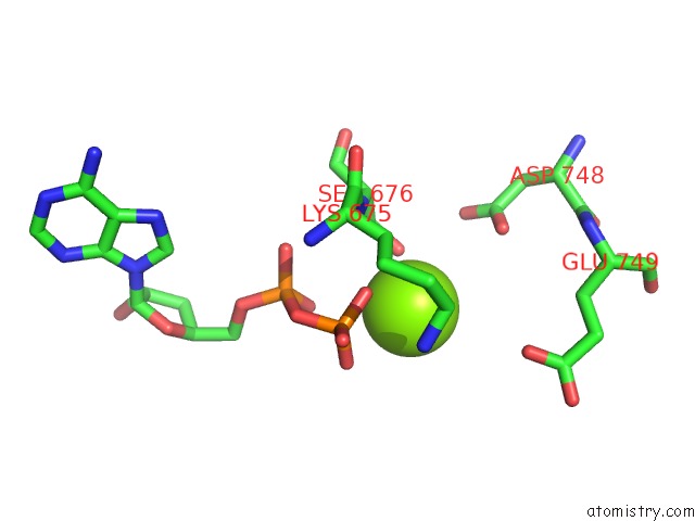
Mono view
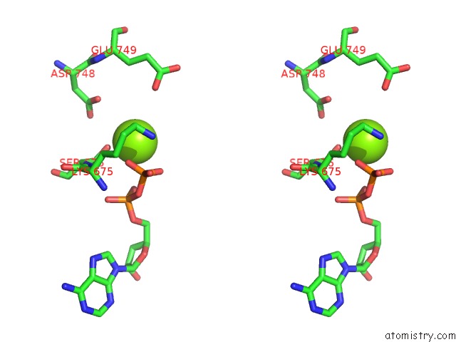
Stereo pair view
Mono view
Stereo pair view
A full contact list of Magnesium with other atoms in the Mg binding
site number 1 of Human Mutsalpha (MSH2/MSH6) Bound to Dna with A Single Base T Insert within 5.0Å range:
|
Reference:
J.J.Warren,
T.J.Pohlhaus,
A.Changela,
R.R.Iyer,
P.L.Modrich,
L.S.Beese.
Structure of the Human Mutsalpha Dna Lesion Recognition Complex. Mol.Cell V. 26 579 2007.
ISSN: ISSN 1097-2765
PubMed: 17531815
DOI: 10.1016/J.MOLCEL.2007.04.018
Page generated: Sun Aug 10 12:24:33 2025
ISSN: ISSN 1097-2765
PubMed: 17531815
DOI: 10.1016/J.MOLCEL.2007.04.018
Last articles
Mg in 6XX7Mg in 6XWD
Mg in 6XW6
Mg in 6XW7
Mg in 6XUP
Mg in 6XUR
Mg in 6XUS
Mg in 6XT7
Mg in 6XU0
Mg in 6XTX