Magnesium »
PDB 2o56-2oh6 »
2oa0 »
Magnesium in PDB 2oa0: Crystal Structure of Calcium Atpase with Bound Adp and Cyclopiazonic Acid
Enzymatic activity of Crystal Structure of Calcium Atpase with Bound Adp and Cyclopiazonic Acid
All present enzymatic activity of Crystal Structure of Calcium Atpase with Bound Adp and Cyclopiazonic Acid:
3.6.3.8;
3.6.3.8;
Protein crystallography data
The structure of Crystal Structure of Calcium Atpase with Bound Adp and Cyclopiazonic Acid, PDB code: 2oa0
was solved by
H.S.Young,
K.A.Moncoq,
with X-Ray Crystallography technique. A brief refinement statistics is given in the table below:
| Resolution Low / High (Å) | 30.00 / 3.40 |
| Space group | P 1 21 1 |
| Cell size a, b, c (Å), α, β, γ (°) | 62.498, 96.836, 154.856, 90.00, 94.83, 90.00 |
| R / Rfree (%) | 29 / 32.8 |
Magnesium Binding Sites:
The binding sites of Magnesium atom in the Crystal Structure of Calcium Atpase with Bound Adp and Cyclopiazonic Acid
(pdb code 2oa0). This binding sites where shown within
5.0 Angstroms radius around Magnesium atom.
In total only one binding site of Magnesium was determined in the Crystal Structure of Calcium Atpase with Bound Adp and Cyclopiazonic Acid, PDB code: 2oa0:
In total only one binding site of Magnesium was determined in the Crystal Structure of Calcium Atpase with Bound Adp and Cyclopiazonic Acid, PDB code: 2oa0:
Magnesium binding site 1 out of 1 in 2oa0
Go back to
Magnesium binding site 1 out
of 1 in the Crystal Structure of Calcium Atpase with Bound Adp and Cyclopiazonic Acid
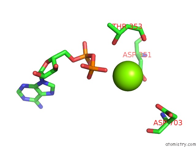
Mono view
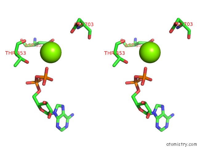
Stereo pair view
Mono view
Stereo pair view
A full contact list of Magnesium with other atoms in the Mg binding
site number 1 of Crystal Structure of Calcium Atpase with Bound Adp and Cyclopiazonic Acid within 5.0Å range:
|
Reference:
K.Moncoq,
C.A.Trieber,
H.S.Young.
The Molecular Basis For Cyclopiazonic Acid Inhibition of the Sarcoplasmic Reticulum Calcium Pump. J.Biol.Chem. V. 282 9748 2007.
ISSN: ISSN 0021-9258
PubMed: 17259168
DOI: 10.1074/JBC.M611653200
Page generated: Sun Aug 10 12:24:48 2025
ISSN: ISSN 0021-9258
PubMed: 17259168
DOI: 10.1074/JBC.M611653200
Last articles
Mg in 5Y8VMg in 5Y88
Mg in 5Y8H
Mg in 5Y8B
Mg in 5Y5P
Mg in 5Y5Q
Mg in 5Y6Z
Mg in 5XYM
Mg in 5XYU
Mg in 5Y4N