Magnesium »
PDB 2p8b-2ppb »
2pan »
Magnesium in PDB 2pan: Crystal Structure of E. Coli Glyoxylate Carboligase
Enzymatic activity of Crystal Structure of E. Coli Glyoxylate Carboligase
All present enzymatic activity of Crystal Structure of E. Coli Glyoxylate Carboligase:
4.1.1.47;
4.1.1.47;
Protein crystallography data
The structure of Crystal Structure of E. Coli Glyoxylate Carboligase, PDB code: 2pan
was solved by
A.Kaplun,
D.M.Chipman,
Z.Barak,
M.Vyazmensky,
B.Shaanan,
with X-Ray Crystallography technique. A brief refinement statistics is given in the table below:
| Resolution Low / High (Å) | 20.00 / 2.70 |
| Space group | P 41 21 2 |
| Cell size a, b, c (Å), α, β, γ (°) | 188.180, 188.180, 249.400, 90.00, 90.00, 90.00 |
| R / Rfree (%) | 21.9 / 25.3 |
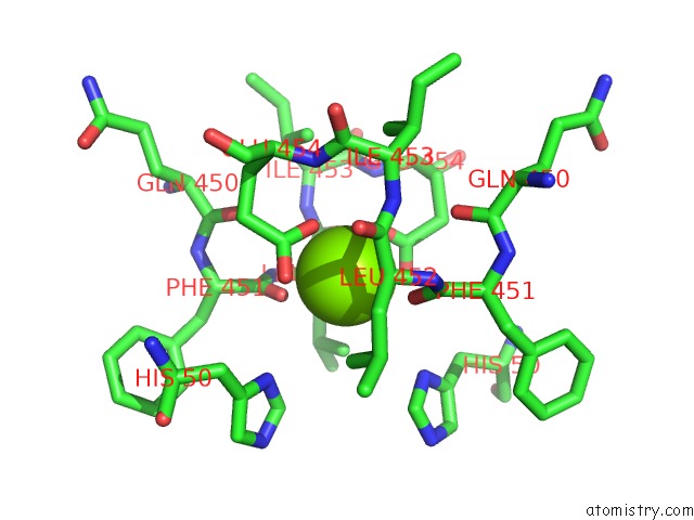
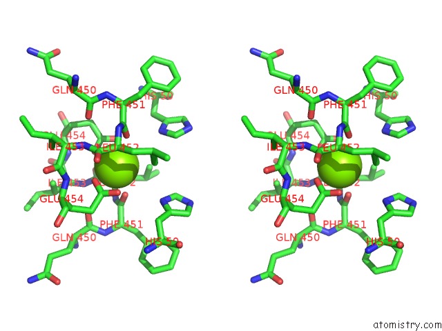
Magnesium Binding Sites:
The binding sites of Magnesium atom in the Crystal Structure of E. Coli Glyoxylate Carboligase
(pdb code 2pan). This binding sites where shown within
5.0 Angstroms radius around Magnesium atom.
In total 10 binding sites of Magnesium where determined in the Crystal Structure of E. Coli Glyoxylate Carboligase, PDB code: 2pan:
Jump to Magnesium binding site number: 1; 2; 3; 4; 5; 6; 7; 8; 9; 10;
In total 10 binding sites of Magnesium where determined in the Crystal Structure of E. Coli Glyoxylate Carboligase, PDB code: 2pan:
Jump to Magnesium binding site number: 1; 2; 3; 4; 5; 6; 7; 8; 9; 10;
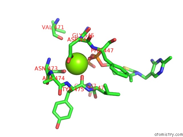
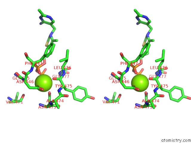
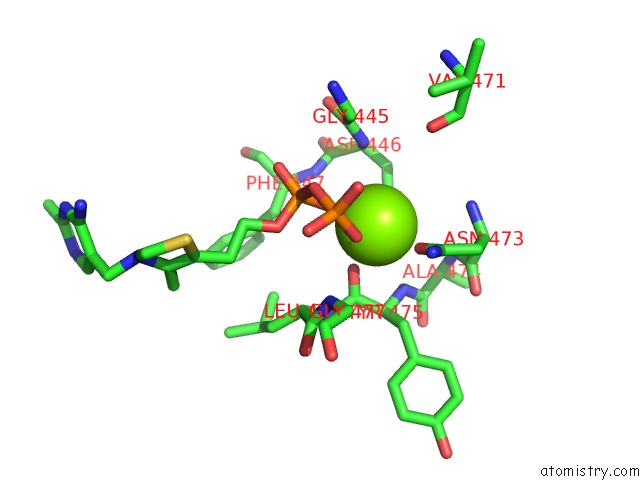
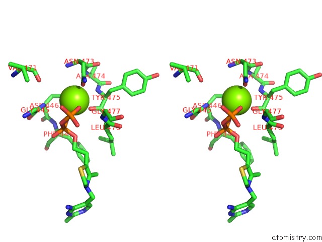
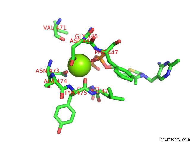
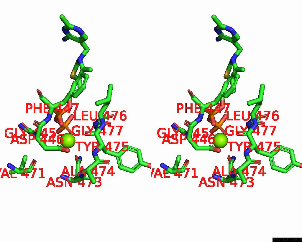
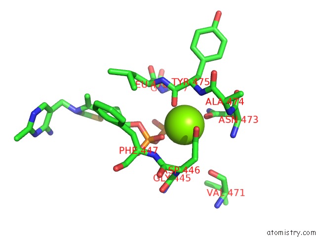
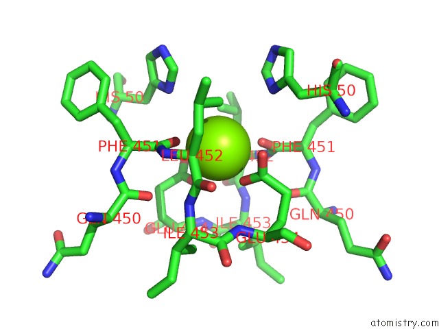
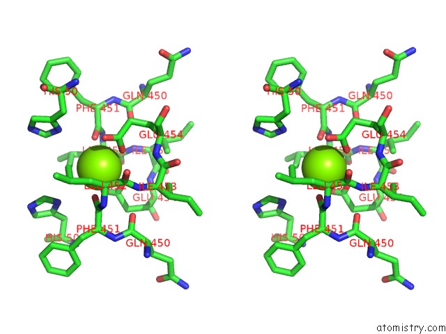
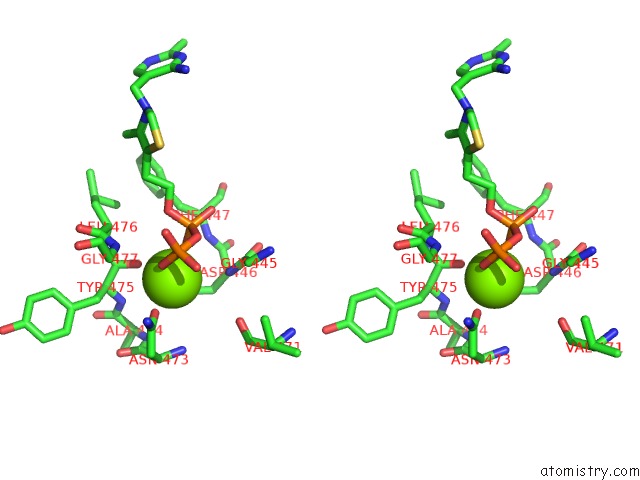
Magnesium binding site 1 out of 10 in 2pan
Go back to
Magnesium binding site 1 out
of 10 in the Crystal Structure of E. Coli Glyoxylate Carboligase
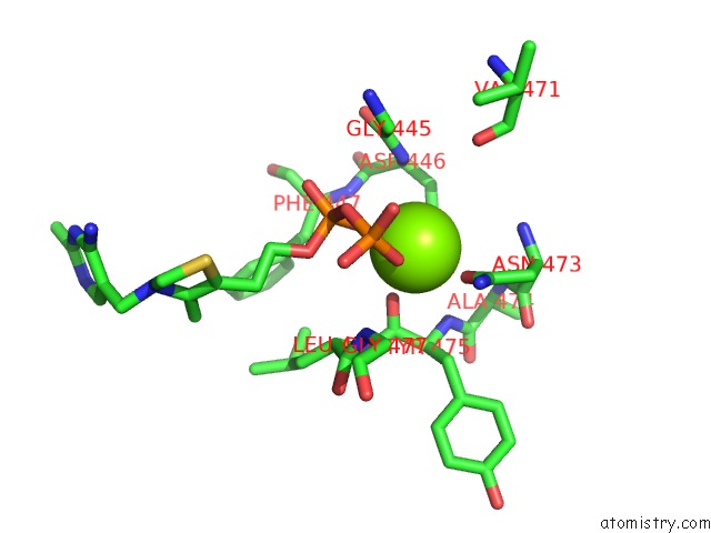
Mono view

Stereo pair view
Mono view
Stereo pair view
A full contact list of Magnesium with other atoms in the Mg binding
site number 1 of Crystal Structure of E. Coli Glyoxylate Carboligase within 5.0Å range:
|
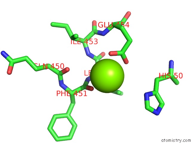
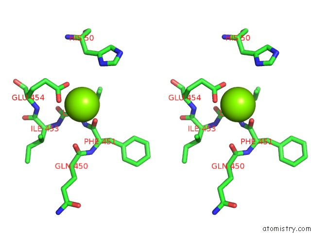
Magnesium binding site 2 out of 10 in 2pan
Go back to
Magnesium binding site 2 out
of 10 in the Crystal Structure of E. Coli Glyoxylate Carboligase
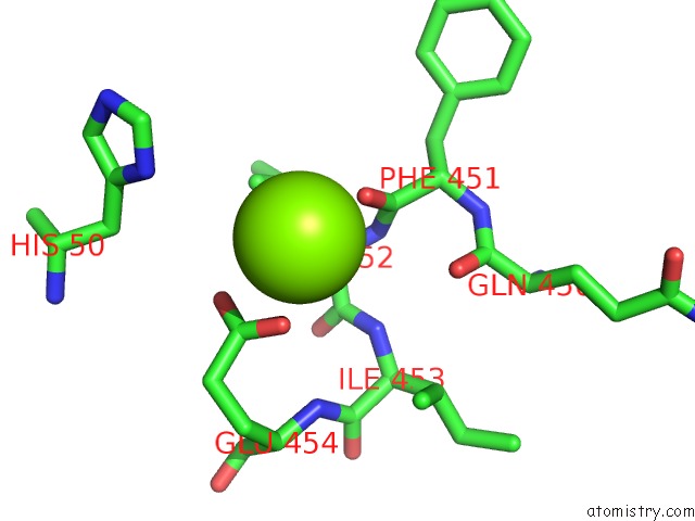
Mono view
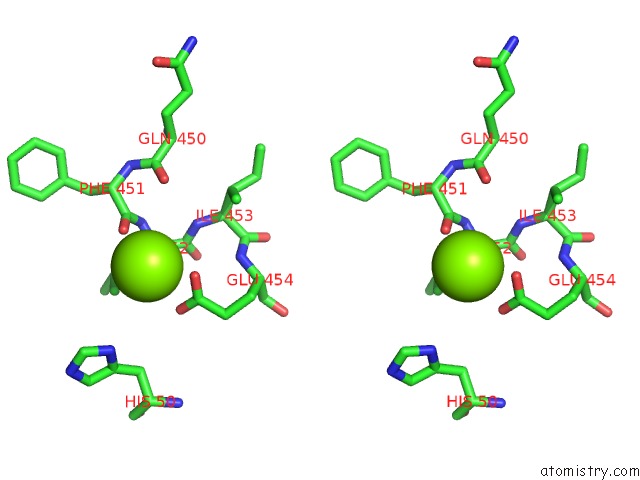
Stereo pair view
Mono view
Stereo pair view
A full contact list of Magnesium with other atoms in the Mg binding
site number 2 of Crystal Structure of E. Coli Glyoxylate Carboligase within 5.0Å range:
|
Magnesium binding site 3 out of 10 in 2pan
Go back to
Magnesium binding site 3 out
of 10 in the Crystal Structure of E. Coli Glyoxylate Carboligase
Mono view
Stereo pair view
Mono view
Stereo pair view
A full contact list of Magnesium with other atoms in the Mg binding
site number 3 of Crystal Structure of E. Coli Glyoxylate Carboligase within 5.0Å range:
|
Magnesium binding site 4 out of 10 in 2pan
Go back to
Magnesium binding site 4 out
of 10 in the Crystal Structure of E. Coli Glyoxylate Carboligase
Mono view
Stereo pair view
Mono view
Stereo pair view
A full contact list of Magnesium with other atoms in the Mg binding
site number 4 of Crystal Structure of E. Coli Glyoxylate Carboligase within 5.0Å range:
|
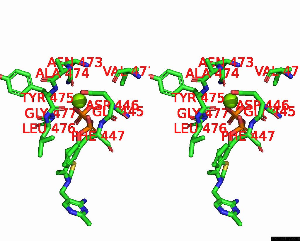
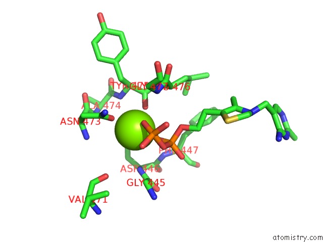
Magnesium binding site 5 out of 10 in 2pan
Go back to
Magnesium binding site 5 out
of 10 in the Crystal Structure of E. Coli Glyoxylate Carboligase
Mono view
Stereo pair view
Mono view
Stereo pair view
A full contact list of Magnesium with other atoms in the Mg binding
site number 5 of Crystal Structure of E. Coli Glyoxylate Carboligase within 5.0Å range:
|
Magnesium binding site 6 out of 10 in 2pan
Go back to
Magnesium binding site 6 out
of 10 in the Crystal Structure of E. Coli Glyoxylate Carboligase
Mono view
Stereo pair view
Mono view
Stereo pair view
A full contact list of Magnesium with other atoms in the Mg binding
site number 6 of Crystal Structure of E. Coli Glyoxylate Carboligase within 5.0Å range:
|
Magnesium binding site 7 out of 10 in 2pan
Go back to
Magnesium binding site 7 out
of 10 in the Crystal Structure of E. Coli Glyoxylate Carboligase
Mono view
Stereo pair view
Mono view
Stereo pair view
A full contact list of Magnesium with other atoms in the Mg binding
site number 7 of Crystal Structure of E. Coli Glyoxylate Carboligase within 5.0Å range:
|
Magnesium binding site 8 out of 10 in 2pan
Go back to
Magnesium binding site 8 out
of 10 in the Crystal Structure of E. Coli Glyoxylate Carboligase
Mono view
Stereo pair view
Mono view
Stereo pair view
A full contact list of Magnesium with other atoms in the Mg binding
site number 8 of Crystal Structure of E. Coli Glyoxylate Carboligase within 5.0Å range:
|
Magnesium binding site 9 out of 10 in 2pan
Go back to
Magnesium binding site 9 out
of 10 in the Crystal Structure of E. Coli Glyoxylate Carboligase
Mono view
Stereo pair view
Mono view
Stereo pair view
A full contact list of Magnesium with other atoms in the Mg binding
site number 9 of Crystal Structure of E. Coli Glyoxylate Carboligase within 5.0Å range:
|
Magnesium binding site 10 out of 10 in 2pan
Go back to
Magnesium binding site 10 out
of 10 in the Crystal Structure of E. Coli Glyoxylate Carboligase
Mono view
Stereo pair view
Mono view
Stereo pair view
A full contact list of Magnesium with other atoms in the Mg binding
site number 10 of Crystal Structure of E. Coli Glyoxylate Carboligase within 5.0Å range:
|
Reference:
A.Kaplun,
E.Binshtein,
M.Vyazmensky,
A.Steinmetz,
Z.Barak,
D.M.Chipman,
K.Tittmann,
B.Shaanan.
Glyoxylate Carboligase Lacks the Canonical Active Site Glutamate of Thiamine-Dependent Enzymes. Nat.Chem.Biol. V. 4 113 2008.
ISSN: ISSN 1552-4450
PubMed: 18176558
DOI: 10.1038/NCHEMBIO.62
Page generated: Sun Aug 10 12:53:30 2025
ISSN: ISSN 1552-4450
PubMed: 18176558
DOI: 10.1038/NCHEMBIO.62
Last articles
Mg in 5WMBMg in 5WM8
Mg in 5WNO
Mg in 5WNI
Mg in 5WMT
Mg in 5WM1
Mg in 5WM6
Mg in 5WM4
Mg in 5WKC
Mg in 5WM3