Magnesium »
PDB 2q9y-2qrf »
2qpl »
Magnesium in PDB 2qpl: Crystal Structure of Calf Spleen Purine Nucleoside Phosphorylase Complexed to A Novel Purine Analogue
Enzymatic activity of Crystal Structure of Calf Spleen Purine Nucleoside Phosphorylase Complexed to A Novel Purine Analogue
All present enzymatic activity of Crystal Structure of Calf Spleen Purine Nucleoside Phosphorylase Complexed to A Novel Purine Analogue:
2.4.2.1;
2.4.2.1;
Protein crystallography data
The structure of Crystal Structure of Calf Spleen Purine Nucleoside Phosphorylase Complexed to A Novel Purine Analogue, PDB code: 2qpl
was solved by
H.M.Pereira,
V.Berdini,
A.Cleasby,
R.C.Garratt,
with X-Ray Crystallography technique. A brief refinement statistics is given in the table below:
| Resolution Low / High (Å) | 46.73 / 2.10 |
| Space group | P 21 3 |
| Cell size a, b, c (Å), α, β, γ (°) | 93.442, 93.442, 93.442, 90.00, 90.00, 90.00 |
| R / Rfree (%) | 16.8 / 21.8 |
Magnesium Binding Sites:
The binding sites of Magnesium atom in the Crystal Structure of Calf Spleen Purine Nucleoside Phosphorylase Complexed to A Novel Purine Analogue
(pdb code 2qpl). This binding sites where shown within
5.0 Angstroms radius around Magnesium atom.
In total only one binding site of Magnesium was determined in the Crystal Structure of Calf Spleen Purine Nucleoside Phosphorylase Complexed to A Novel Purine Analogue, PDB code: 2qpl:
In total only one binding site of Magnesium was determined in the Crystal Structure of Calf Spleen Purine Nucleoside Phosphorylase Complexed to A Novel Purine Analogue, PDB code: 2qpl:
Magnesium binding site 1 out of 1 in 2qpl
Go back to
Magnesium binding site 1 out
of 1 in the Crystal Structure of Calf Spleen Purine Nucleoside Phosphorylase Complexed to A Novel Purine Analogue
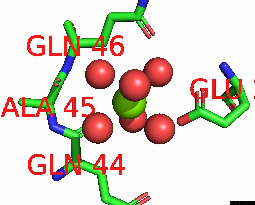
Mono view
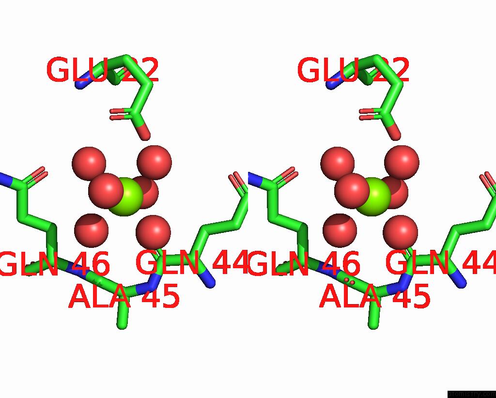
Stereo pair view
Mono view
Stereo pair view
A full contact list of Magnesium with other atoms in the Mg binding
site number 1 of Crystal Structure of Calf Spleen Purine Nucleoside Phosphorylase Complexed to A Novel Purine Analogue within 5.0Å range:
|
Reference:
H.M.Pereira,
V.Berdini,
A.Cleasby,
R.C.Garratt.
Crystal Structure of Calf Spleen Purine Nucleoside Phosphorylase Complexed to A Novel Purine Analogue. Febs Lett. V. 581 5082 2007.
ISSN: ISSN 0014-5793
PubMed: 17927987
DOI: 10.1016/J.FEBSLET.2007.09.051
Page generated: Sun Aug 10 13:25:27 2025
ISSN: ISSN 0014-5793
PubMed: 17927987
DOI: 10.1016/J.FEBSLET.2007.09.051
Last articles
Mg in 3T99Mg in 3T8V
Mg in 3T80
Mg in 3T5P
Mg in 3T8Q
Mg in 3T8O
Mg in 3T7A
Mg in 3T77
Mg in 3T6E
Mg in 3T6R