Magnesium »
PDB 2xbp-2xnd »
2xh2 »
Magnesium in PDB 2xh2: Engineering the Enolase Active Site Pocket: Crystal Structure of the S39N D321A Mutant of Yeast Enolase 1
Enzymatic activity of Engineering the Enolase Active Site Pocket: Crystal Structure of the S39N D321A Mutant of Yeast Enolase 1
All present enzymatic activity of Engineering the Enolase Active Site Pocket: Crystal Structure of the S39N D321A Mutant of Yeast Enolase 1:
4.2.1.11;
4.2.1.11;
Protein crystallography data
The structure of Engineering the Enolase Active Site Pocket: Crystal Structure of the S39N D321A Mutant of Yeast Enolase 1, PDB code: 2xh2
was solved by
B.Schreier,
B.Hocker,
with X-Ray Crystallography technique. A brief refinement statistics is given in the table below:
| Resolution Low / High (Å) | 35.65 / 1.80 |
| Space group | P 1 |
| Cell size a, b, c (Å), α, β, γ (°) | 64.720, 82.540, 95.610, 89.41, 71.31, 84.80 |
| R / Rfree (%) | 16.579 / 20.949 |
Magnesium Binding Sites:
The binding sites of Magnesium atom in the Engineering the Enolase Active Site Pocket: Crystal Structure of the S39N D321A Mutant of Yeast Enolase 1
(pdb code 2xh2). This binding sites where shown within
5.0 Angstroms radius around Magnesium atom.
In total 4 binding sites of Magnesium where determined in the Engineering the Enolase Active Site Pocket: Crystal Structure of the S39N D321A Mutant of Yeast Enolase 1, PDB code: 2xh2:
Jump to Magnesium binding site number: 1; 2; 3; 4;
In total 4 binding sites of Magnesium where determined in the Engineering the Enolase Active Site Pocket: Crystal Structure of the S39N D321A Mutant of Yeast Enolase 1, PDB code: 2xh2:
Jump to Magnesium binding site number: 1; 2; 3; 4;
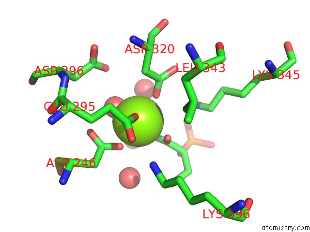
Magnesium binding site 1 out of 4 in 2xh2
Go back to
Magnesium binding site 1 out
of 4 in the Engineering the Enolase Active Site Pocket: Crystal Structure of the S39N D321A Mutant of Yeast Enolase 1
Mono view

Stereo pair view
Mono view
Stereo pair view
A full contact list of Magnesium with other atoms in the Mg binding
site number 1 of Engineering the Enolase Active Site Pocket: Crystal Structure of the S39N D321A Mutant of Yeast Enolase 1 within 5.0Å range:
|
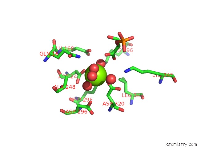
Magnesium binding site 2 out of 4 in 2xh2
Go back to
Magnesium binding site 2 out
of 4 in the Engineering the Enolase Active Site Pocket: Crystal Structure of the S39N D321A Mutant of Yeast Enolase 1
Mono view

Stereo pair view
Mono view
Stereo pair view
A full contact list of Magnesium with other atoms in the Mg binding
site number 2 of Engineering the Enolase Active Site Pocket: Crystal Structure of the S39N D321A Mutant of Yeast Enolase 1 within 5.0Å range:
|
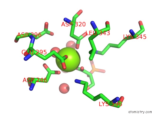
Magnesium binding site 3 out of 4 in 2xh2
Go back to
Magnesium binding site 3 out
of 4 in the Engineering the Enolase Active Site Pocket: Crystal Structure of the S39N D321A Mutant of Yeast Enolase 1
Mono view

Stereo pair view
Mono view
Stereo pair view
A full contact list of Magnesium with other atoms in the Mg binding
site number 3 of Engineering the Enolase Active Site Pocket: Crystal Structure of the S39N D321A Mutant of Yeast Enolase 1 within 5.0Å range:
|
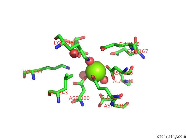
Magnesium binding site 4 out of 4 in 2xh2
Go back to
Magnesium binding site 4 out
of 4 in the Engineering the Enolase Active Site Pocket: Crystal Structure of the S39N D321A Mutant of Yeast Enolase 1
Mono view

Stereo pair view
Mono view
Stereo pair view
A full contact list of Magnesium with other atoms in the Mg binding
site number 4 of Engineering the Enolase Active Site Pocket: Crystal Structure of the S39N D321A Mutant of Yeast Enolase 1 within 5.0Å range:
|
Reference:
B.Schreier,
B.Hoecker.
Engineering the Enolase Magnesium II Binding Site - Implications For Its Evolution. Biochemistry V. 49 7582 2010.
ISSN: ISSN 0006-2960
PubMed: 20690637
DOI: 10.1021/BI100954F
Page generated: Sun Aug 10 16:25:05 2025
ISSN: ISSN 0006-2960
PubMed: 20690637
DOI: 10.1021/BI100954F
Last articles
Mg in 6DCFMg in 6DA6
Mg in 6D9S
Mg in 6DB9
Mg in 6DBY
Mg in 6D9R
Mg in 6D9L
Mg in 6D95
Mg in 6D9K
Mg in 6D9M