Magnesium »
PDB 3a0u-3abk »
3a7r »
Magnesium in PDB 3a7r: Crystal Structure of E. Coli Lipoate-Protein Ligase A in Complex with Lipoyl-Amp.
Protein crystallography data
The structure of Crystal Structure of E. Coli Lipoate-Protein Ligase A in Complex with Lipoyl-Amp., PDB code: 3a7r
was solved by
K.Fujiwara,
H.Hosaka,
with X-Ray Crystallography technique. A brief refinement statistics is given in the table below:
| Resolution Low / High (Å) | 20.00 / 2.05 |
| Space group | P 61 2 2 |
| Cell size a, b, c (Å), α, β, γ (°) | 102.298, 102.298, 221.252, 90.00, 90.00, 120.00 |
| R / Rfree (%) | 17.6 / 20.4 |
Magnesium Binding Sites:
The binding sites of Magnesium atom in the Crystal Structure of E. Coli Lipoate-Protein Ligase A in Complex with Lipoyl-Amp.
(pdb code 3a7r). This binding sites where shown within
5.0 Angstroms radius around Magnesium atom.
In total only one binding site of Magnesium was determined in the Crystal Structure of E. Coli Lipoate-Protein Ligase A in Complex with Lipoyl-Amp., PDB code: 3a7r:
In total only one binding site of Magnesium was determined in the Crystal Structure of E. Coli Lipoate-Protein Ligase A in Complex with Lipoyl-Amp., PDB code: 3a7r:
Magnesium binding site 1 out of 1 in 3a7r
Go back to
Magnesium binding site 1 out
of 1 in the Crystal Structure of E. Coli Lipoate-Protein Ligase A in Complex with Lipoyl-Amp.
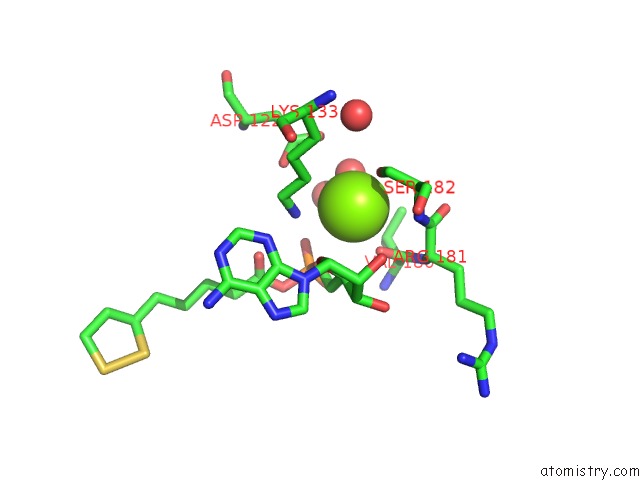
Mono view
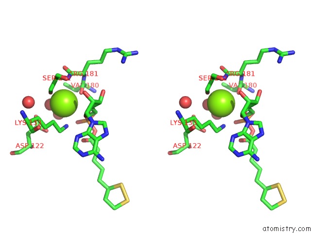
Stereo pair view
Mono view
Stereo pair view
A full contact list of Magnesium with other atoms in the Mg binding
site number 1 of Crystal Structure of E. Coli Lipoate-Protein Ligase A in Complex with Lipoyl-Amp. within 5.0Å range:
|
Reference:
K.Fujiwara,
N.Maita,
H.Hosaka,
K.Okamura-Ikeda,
A.Nakagawa,
H.Taniguchi.
Global Conformational Change Associated with the Two-Step Reaction Catalyzed By Escherichia Coli Lipoate-Protein Ligase A. J.Biol.Chem. V. 285 9971 2010.
ISSN: ISSN 0021-9258
PubMed: 20089862
DOI: 10.1074/JBC.M109.078717
Page generated: Sun Aug 10 17:23:37 2025
ISSN: ISSN 0021-9258
PubMed: 20089862
DOI: 10.1074/JBC.M109.078717
Last articles
Mg in 4ZK5Mg in 4ZJJ
Mg in 4ZJI
Mg in 4ZK4
Mg in 4ZIR
Mg in 4ZIY
Mg in 4ZIB
Mg in 4ZI7
Mg in 4ZIL
Mg in 4ZI5