Magnesium »
PDB 3abl-3aln »
3ajk »
Magnesium in PDB 3ajk: Crystal Structure of D(CGCGGATF5UCGCG): 5-Formyluridine:Guanosine Base-Pair in B-Dna with HOECHST33258
Protein crystallography data
The structure of Crystal Structure of D(CGCGGATF5UCGCG): 5-Formyluridine:Guanosine Base-Pair in B-Dna with HOECHST33258, PDB code: 3ajk
was solved by
M.Tsunoda,
T.Sakaue,
Y.Ueno,
A.Matsuda,
A.Takenaka,
with X-Ray Crystallography technique. A brief refinement statistics is given in the table below:
| Resolution Low / High (Å) | 10.00 / 1.95 |
| Space group | P 21 21 21 |
| Cell size a, b, c (Å), α, β, γ (°) | 25.327, 40.331, 65.961, 90.00, 90.00, 90.00 |
| R / Rfree (%) | 20.3 / 26.1 |
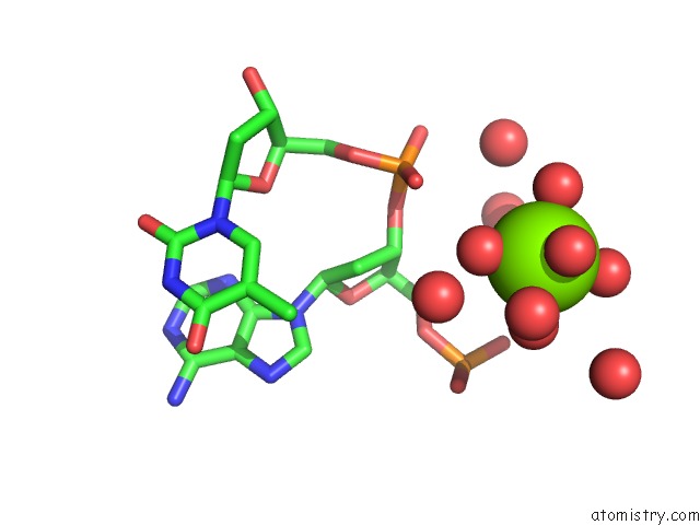
Magnesium Binding Sites:
The binding sites of Magnesium atom in the Crystal Structure of D(CGCGGATF5UCGCG): 5-Formyluridine:Guanosine Base-Pair in B-Dna with HOECHST33258
(pdb code 3ajk). This binding sites where shown within
5.0 Angstroms radius around Magnesium atom.
In total only one binding site of Magnesium was determined in the Crystal Structure of D(CGCGGATF5UCGCG): 5-Formyluridine:Guanosine Base-Pair in B-Dna with HOECHST33258, PDB code: 3ajk:
In total only one binding site of Magnesium was determined in the Crystal Structure of D(CGCGGATF5UCGCG): 5-Formyluridine:Guanosine Base-Pair in B-Dna with HOECHST33258, PDB code: 3ajk:
Magnesium binding site 1 out of 1 in 3ajk
Go back to
Magnesium binding site 1 out
of 1 in the Crystal Structure of D(CGCGGATF5UCGCG): 5-Formyluridine:Guanosine Base-Pair in B-Dna with HOECHST33258
Mono view
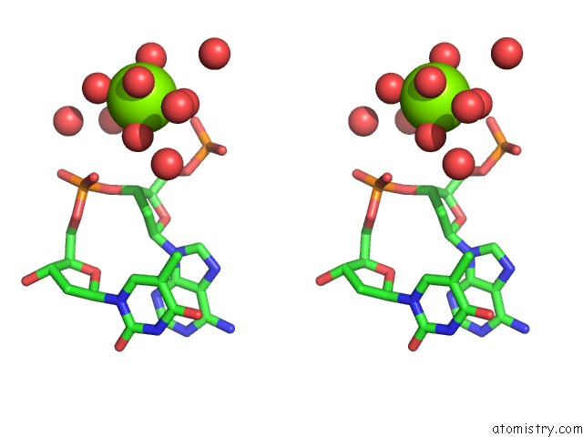
Stereo pair view
Mono view
Stereo pair view
A full contact list of Magnesium with other atoms in the Mg binding
site number 1 of Crystal Structure of D(CGCGGATF5UCGCG): 5-Formyluridine:Guanosine Base-Pair in B-Dna with HOECHST33258 within 5.0Å range:
|
Reference:
M.Tsunoda,
T.Sakaue,
S.Naito,
T.Sunami,
N.Abe,
Y.Ueno,
A.Matsuda,
A.Takenaka.
Insights Into the Structures of Dna Damaged By Hydroxyl Radical: Crystal Structures of Dna Duplexes Containing 5-Formyluracil J Nucleic Acids V.2010 10728 2010.
ISSN: ISSN 2090-0201
PubMed: 20976303
DOI: 10.4061/2010/107289
Page generated: Sun Aug 10 17:27:46 2025
ISSN: ISSN 2090-0201
PubMed: 20976303
DOI: 10.4061/2010/107289
Last articles
Mg in 5WMBMg in 5WM8
Mg in 5WNO
Mg in 5WNI
Mg in 5WMT
Mg in 5WM1
Mg in 5WM6
Mg in 5WM4
Mg in 5WKC
Mg in 5WM3