Magnesium »
PDB 3e3z-3ef8 »
3e4c »
Magnesium in PDB 3e4c: Procaspase-1 Zymogen Domain Crystal Strucutre
Enzymatic activity of Procaspase-1 Zymogen Domain Crystal Strucutre
All present enzymatic activity of Procaspase-1 Zymogen Domain Crystal Strucutre:
3.4.22.36;
3.4.22.36;
Protein crystallography data
The structure of Procaspase-1 Zymogen Domain Crystal Strucutre, PDB code: 3e4c
was solved by
J.M.Elliott,
L.Rouge,
C.Wiesmann,
J.M.Scheer,
with X-Ray Crystallography technique. A brief refinement statistics is given in the table below:
| Resolution Low / High (Å) | 20.00 / 2.05 |
| Space group | P 1 |
| Cell size a, b, c (Å), α, β, γ (°) | 56.100, 58.004, 60.120, 105.95, 119.60, 92.77 |
| R / Rfree (%) | 20 / 26.3 |
Magnesium Binding Sites:
The binding sites of Magnesium atom in the Procaspase-1 Zymogen Domain Crystal Strucutre
(pdb code 3e4c). This binding sites where shown within
5.0 Angstroms radius around Magnesium atom.
In total only one binding site of Magnesium was determined in the Procaspase-1 Zymogen Domain Crystal Strucutre, PDB code: 3e4c:
In total only one binding site of Magnesium was determined in the Procaspase-1 Zymogen Domain Crystal Strucutre, PDB code: 3e4c:
Magnesium binding site 1 out of 1 in 3e4c
Go back to
Magnesium binding site 1 out
of 1 in the Procaspase-1 Zymogen Domain Crystal Strucutre
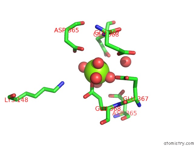
Mono view
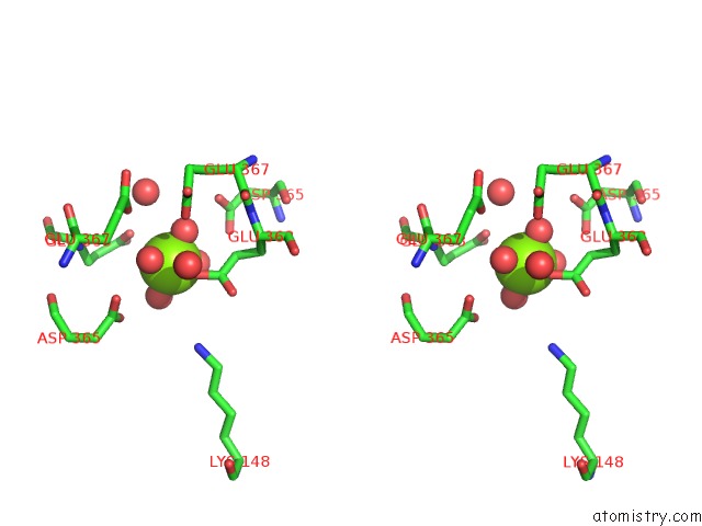
Stereo pair view
Mono view
Stereo pair view
A full contact list of Magnesium with other atoms in the Mg binding
site number 1 of Procaspase-1 Zymogen Domain Crystal Strucutre within 5.0Å range:
|
Reference:
J.M.Elliott,
L.Rouge,
C.Wiesmann,
J.M.Scheer.
Crystal Structure of Procaspase-1 Zymogen Domain Reveals Insight Into Inflammatory Caspase Autoactivation J.Biol.Chem. V. 284 6546 2009.
ISSN: ISSN 0021-9258
PubMed: 19117953
DOI: 10.1074/JBC.M806121200
Page generated: Sun Aug 10 20:23:09 2025
ISSN: ISSN 0021-9258
PubMed: 19117953
DOI: 10.1074/JBC.M806121200
Last articles
Mg in 6EVWMg in 6EVY
Mg in 6EVX
Mg in 6EU3
Mg in 6EU2
Mg in 6EPZ
Mg in 6EU1
Mg in 6EU0
Mg in 6ETH
Mg in 6ETF