Magnesium »
PDB 3hx0-3i5x »
3hy2 »
Magnesium in PDB 3hy2: Crystal Structure of Sulfiredoxin in Complex with Peroxiredoxin I and Atp:MG2+
Enzymatic activity of Crystal Structure of Sulfiredoxin in Complex with Peroxiredoxin I and Atp:MG2+
All present enzymatic activity of Crystal Structure of Sulfiredoxin in Complex with Peroxiredoxin I and Atp:MG2+:
1.11.1.15; 1.8.98.2;
1.11.1.15; 1.8.98.2;
Protein crystallography data
The structure of Crystal Structure of Sulfiredoxin in Complex with Peroxiredoxin I and Atp:MG2+, PDB code: 3hy2
was solved by
T.J.Jonsson,
L.C.Johnson,
W.T.Lowther,
with X-Ray Crystallography technique. A brief refinement statistics is given in the table below:
| Resolution Low / High (Å) | 27.38 / 2.10 |
| Space group | P 21 21 21 |
| Cell size a, b, c (Å), α, β, γ (°) | 57.330, 92.410, 131.850, 90.00, 90.00, 90.00 |
| R / Rfree (%) | 22.4 / 27.3 |
Magnesium Binding Sites:
The binding sites of Magnesium atom in the Crystal Structure of Sulfiredoxin in Complex with Peroxiredoxin I and Atp:MG2+
(pdb code 3hy2). This binding sites where shown within
5.0 Angstroms radius around Magnesium atom.
In total 2 binding sites of Magnesium where determined in the Crystal Structure of Sulfiredoxin in Complex with Peroxiredoxin I and Atp:MG2+, PDB code: 3hy2:
Jump to Magnesium binding site number: 1; 2;
In total 2 binding sites of Magnesium where determined in the Crystal Structure of Sulfiredoxin in Complex with Peroxiredoxin I and Atp:MG2+, PDB code: 3hy2:
Jump to Magnesium binding site number: 1; 2;
Magnesium binding site 1 out of 2 in 3hy2
Go back to
Magnesium binding site 1 out
of 2 in the Crystal Structure of Sulfiredoxin in Complex with Peroxiredoxin I and Atp:MG2+
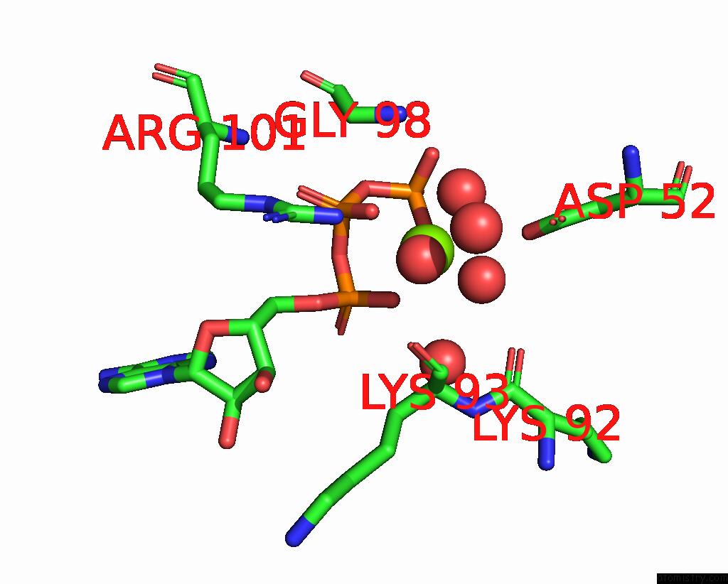
Mono view
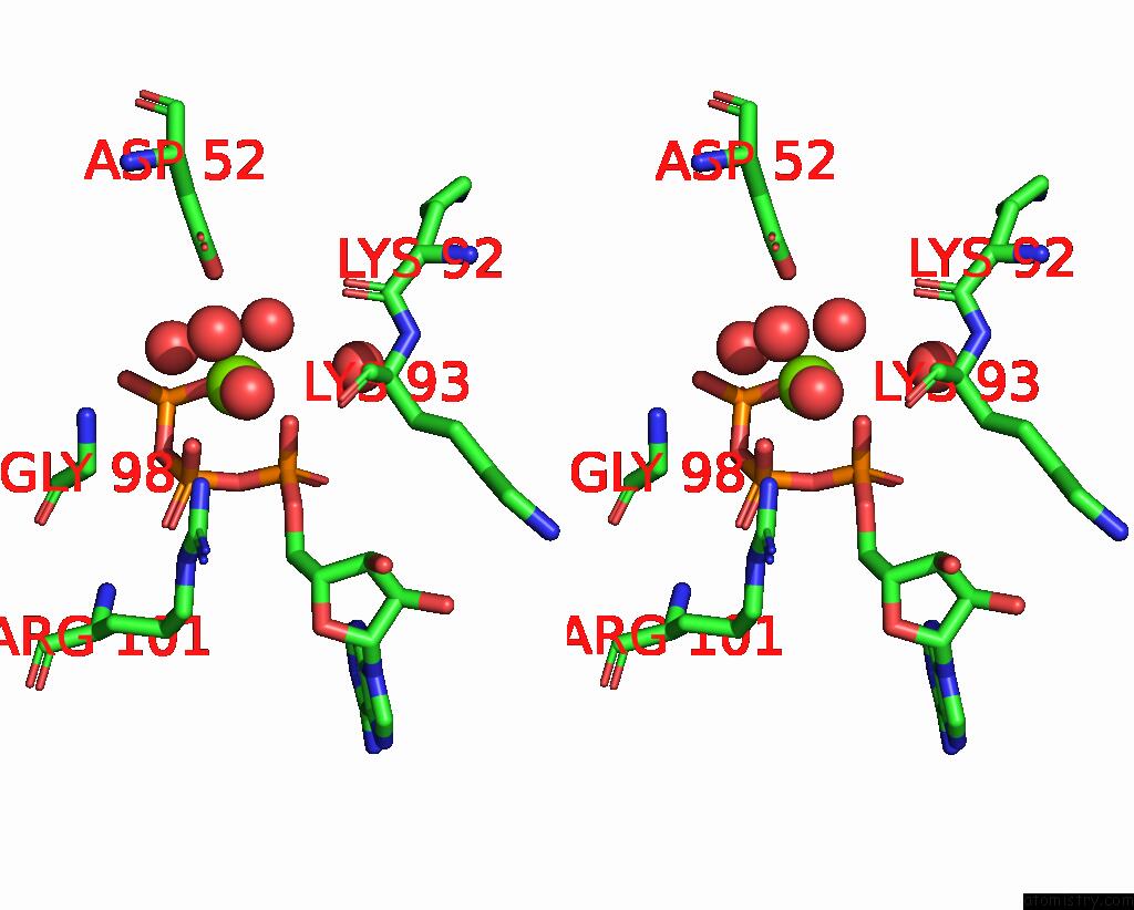
Stereo pair view
Mono view
Stereo pair view
A full contact list of Magnesium with other atoms in the Mg binding
site number 1 of Crystal Structure of Sulfiredoxin in Complex with Peroxiredoxin I and Atp:MG2+ within 5.0Å range:
|
Magnesium binding site 2 out of 2 in 3hy2
Go back to
Magnesium binding site 2 out
of 2 in the Crystal Structure of Sulfiredoxin in Complex with Peroxiredoxin I and Atp:MG2+
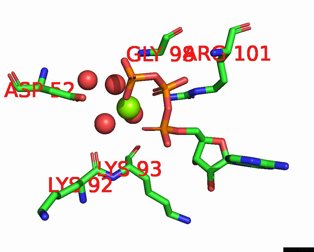
Mono view
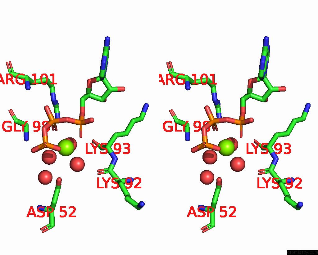
Stereo pair view
Mono view
Stereo pair view
A full contact list of Magnesium with other atoms in the Mg binding
site number 2 of Crystal Structure of Sulfiredoxin in Complex with Peroxiredoxin I and Atp:MG2+ within 5.0Å range:
|
Reference:
T.J.Jonsson,
L.C.Johnson,
W.T.Lowther.
Protein Engineering of the Quaternary Sulfiredoxin-Peroxiredoxin Enzyme-Substrate Complex Reveals the Molecular Basis For Cysteine Sulfinic Acid Phosphorylation J.Biol.Chem. V. 284 33305 2009.
ISSN: ISSN 0021-9258
PubMed: 19812042
DOI: 10.1074/JBC.M109.036400
Page generated: Sun Aug 10 22:10:00 2025
ISSN: ISSN 0021-9258
PubMed: 19812042
DOI: 10.1074/JBC.M109.036400
Last articles
Mg in 3TWAMg in 3TW0
Mg in 3TW7
Mg in 3TVY
Mg in 3TVX
Mg in 3TVD
Mg in 3TTZ
Mg in 3TVK
Mg in 3TVB
Mg in 3TVA