Magnesium »
PDB 3hx0-3i5x »
3hyo »
Magnesium in PDB 3hyo: Crystal Structure of Pyridoxal Kinase From Lactobacillus Plantarum in Complex with Adp
Enzymatic activity of Crystal Structure of Pyridoxal Kinase From Lactobacillus Plantarum in Complex with Adp
All present enzymatic activity of Crystal Structure of Pyridoxal Kinase From Lactobacillus Plantarum in Complex with Adp:
2.7.1.35;
2.7.1.35;
Protein crystallography data
The structure of Crystal Structure of Pyridoxal Kinase From Lactobacillus Plantarum in Complex with Adp, PDB code: 3hyo
was solved by
A.Bagaria,
D.Kumaran,
S.K.Burley,
S.Swaminathan,
New York Sgx Researchcenter For Structural Genomics (Nysgxrc),
with X-Ray Crystallography technique. A brief refinement statistics is given in the table below:
| Resolution Low / High (Å) | 24.06 / 1.85 |
| Space group | C 2 2 21 |
| Cell size a, b, c (Å), α, β, γ (°) | 64.594, 69.761, 132.924, 90.00, 90.00, 90.00 |
| R / Rfree (%) | 18 / 22.2 |
Magnesium Binding Sites:
The binding sites of Magnesium atom in the Crystal Structure of Pyridoxal Kinase From Lactobacillus Plantarum in Complex with Adp
(pdb code 3hyo). This binding sites where shown within
5.0 Angstroms radius around Magnesium atom.
In total only one binding site of Magnesium was determined in the Crystal Structure of Pyridoxal Kinase From Lactobacillus Plantarum in Complex with Adp, PDB code: 3hyo:
In total only one binding site of Magnesium was determined in the Crystal Structure of Pyridoxal Kinase From Lactobacillus Plantarum in Complex with Adp, PDB code: 3hyo:
Magnesium binding site 1 out of 1 in 3hyo
Go back to
Magnesium binding site 1 out
of 1 in the Crystal Structure of Pyridoxal Kinase From Lactobacillus Plantarum in Complex with Adp
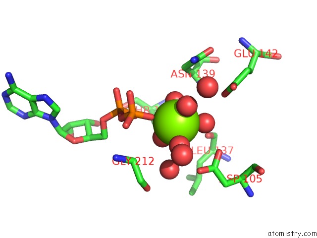
Mono view
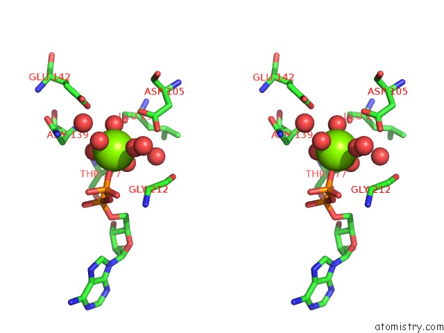
Stereo pair view
Mono view
Stereo pair view
A full contact list of Magnesium with other atoms in the Mg binding
site number 1 of Crystal Structure of Pyridoxal Kinase From Lactobacillus Plantarum in Complex with Adp within 5.0Å range:
|
Reference:
A.Bagaria,
D.Kumaran,
S.K.Burley,
S.Swaminathan.
Crystal Structure of Pyridoxal Kinase From Lactobacillus Plantarum in Complex with Adp To Be Published.
Page generated: Sun Aug 10 22:12:13 2025
Last articles
Mg in 4FR8Mg in 4FS1
Mg in 4FRZ
Mg in 4FQF
Mg in 4FR3
Mg in 4FPV
Mg in 4FNJ
Mg in 4FPP
Mg in 4FP1
Mg in 4FO6