Magnesium »
PDB 3hx0-3i5x »
3i2b »
Magnesium in PDB 3i2b: The Crystal Structure of Human 6 Pyruvoyl Tetrahydrobiopterin Synthase
Enzymatic activity of The Crystal Structure of Human 6 Pyruvoyl Tetrahydrobiopterin Synthase
All present enzymatic activity of The Crystal Structure of Human 6 Pyruvoyl Tetrahydrobiopterin Synthase:
4.2.3.12;
4.2.3.12;
Protein crystallography data
The structure of The Crystal Structure of Human 6 Pyruvoyl Tetrahydrobiopterin Synthase, PDB code: 3i2b
was solved by
E.Ugochukwu,
R.Cocking,
E.Pilka,
W.W.Yue,
J.E.Bray,
A.Chaikuad,
T.Krojer,
J.Muniz,
F.Von Delft,
C.Bountra,
C.H.Arrowsmith,
J.Weigelt,
A.Edwards,
U.Oppermann,
Structural Genomics Consortium (Sgc),
with X-Ray Crystallography technique. A brief refinement statistics is given in the table below:
| Resolution Low / High (Å) | 42.94 / 2.30 |
| Space group | P 21 21 21 |
| Cell size a, b, c (Å), α, β, γ (°) | 73.290, 118.720, 234.980, 90.00, 90.00, 90.00 |
| R / Rfree (%) | 20.6 / 24.6 |
Other elements in 3i2b:
The structure of The Crystal Structure of Human 6 Pyruvoyl Tetrahydrobiopterin Synthase also contains other interesting chemical elements:
| Nickel | (Ni) | 12 atoms |
Magnesium Binding Sites:
The binding sites of Magnesium atom in the The Crystal Structure of Human 6 Pyruvoyl Tetrahydrobiopterin Synthase
(pdb code 3i2b). This binding sites where shown within
5.0 Angstroms radius around Magnesium atom.
In total 2 binding sites of Magnesium where determined in the The Crystal Structure of Human 6 Pyruvoyl Tetrahydrobiopterin Synthase, PDB code: 3i2b:
Jump to Magnesium binding site number: 1; 2;
In total 2 binding sites of Magnesium where determined in the The Crystal Structure of Human 6 Pyruvoyl Tetrahydrobiopterin Synthase, PDB code: 3i2b:
Jump to Magnesium binding site number: 1; 2;
Magnesium binding site 1 out of 2 in 3i2b
Go back to
Magnesium binding site 1 out
of 2 in the The Crystal Structure of Human 6 Pyruvoyl Tetrahydrobiopterin Synthase
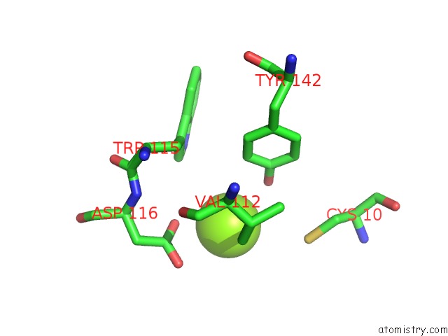
Mono view
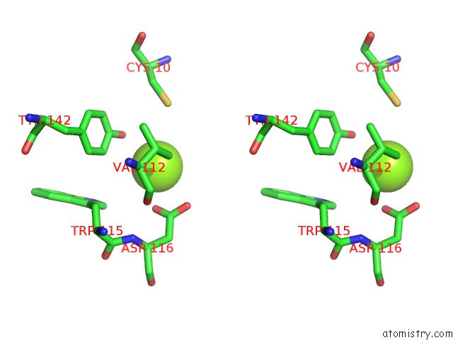
Stereo pair view
Mono view
Stereo pair view
A full contact list of Magnesium with other atoms in the Mg binding
site number 1 of The Crystal Structure of Human 6 Pyruvoyl Tetrahydrobiopterin Synthase within 5.0Å range:
|
Magnesium binding site 2 out of 2 in 3i2b
Go back to
Magnesium binding site 2 out
of 2 in the The Crystal Structure of Human 6 Pyruvoyl Tetrahydrobiopterin Synthase
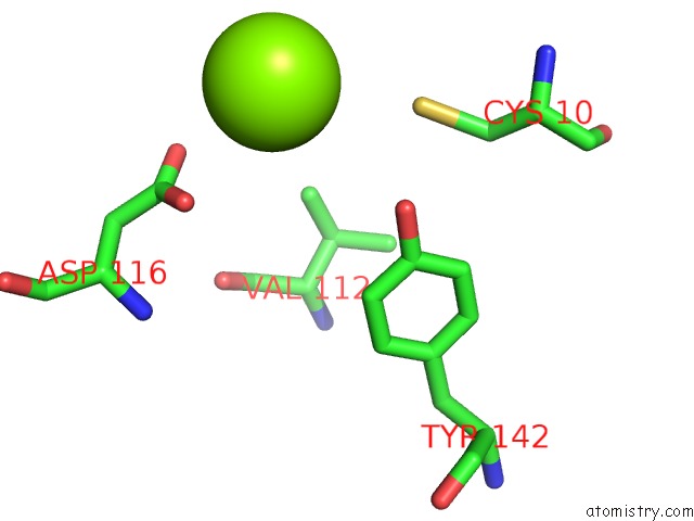
Mono view
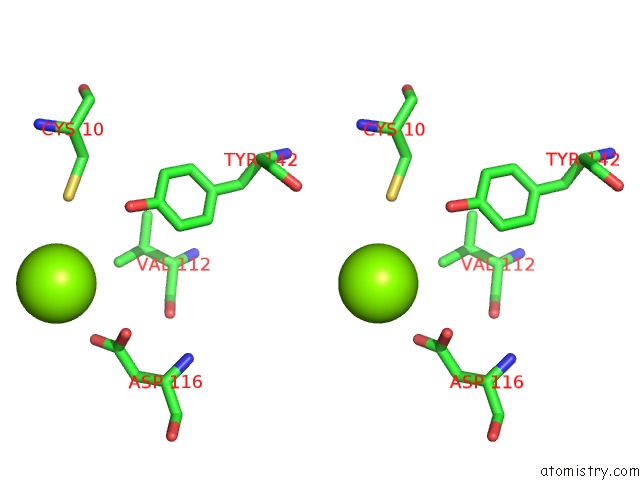
Stereo pair view
Mono view
Stereo pair view
A full contact list of Magnesium with other atoms in the Mg binding
site number 2 of The Crystal Structure of Human 6 Pyruvoyl Tetrahydrobiopterin Synthase within 5.0Å range:
|
Reference:
E.Ugochukwu,
R.Cocking,
E.Pilka,
W.W.Yue,
J.E.Bray,
A.Chaikuad,
T.Krojer,
J.Muniz,
U.Oppermann.
The Crystal Structure of Human 6 Pyruvoyl Tetrahydrobiopterin Synthase To Be Published.
Page generated: Sun Aug 10 22:13:14 2025
Last articles
Mg in 3TWPMg in 3TWB
Mg in 3TWH
Mg in 3TW6
Mg in 3TWA
Mg in 3TW0
Mg in 3TW7
Mg in 3TVY
Mg in 3TVX
Mg in 3TVD