Magnesium »
PDB 3igi-3is5 »
3ijs »
Magnesium in PDB 3ijs: Structure of S67-27 in Complex with Tsbp
Protein crystallography data
The structure of Structure of S67-27 in Complex with Tsbp, PDB code: 3ijs
was solved by
C.L.Brooks,
R.J.Blackler,
S.V.Evans,
with X-Ray Crystallography technique. A brief refinement statistics is given in the table below:
| Resolution Low / High (Å) | 19.93 / 2.55 |
| Space group | P 21 21 21 |
| Cell size a, b, c (Å), α, β, γ (°) | 46.320, 127.910, 156.460, 90.00, 90.00, 90.00 |
| R / Rfree (%) | 21.7 / 26.1 |
Magnesium Binding Sites:
The binding sites of Magnesium atom in the Structure of S67-27 in Complex with Tsbp
(pdb code 3ijs). This binding sites where shown within
5.0 Angstroms radius around Magnesium atom.
In total 2 binding sites of Magnesium where determined in the Structure of S67-27 in Complex with Tsbp, PDB code: 3ijs:
Jump to Magnesium binding site number: 1; 2;
In total 2 binding sites of Magnesium where determined in the Structure of S67-27 in Complex with Tsbp, PDB code: 3ijs:
Jump to Magnesium binding site number: 1; 2;
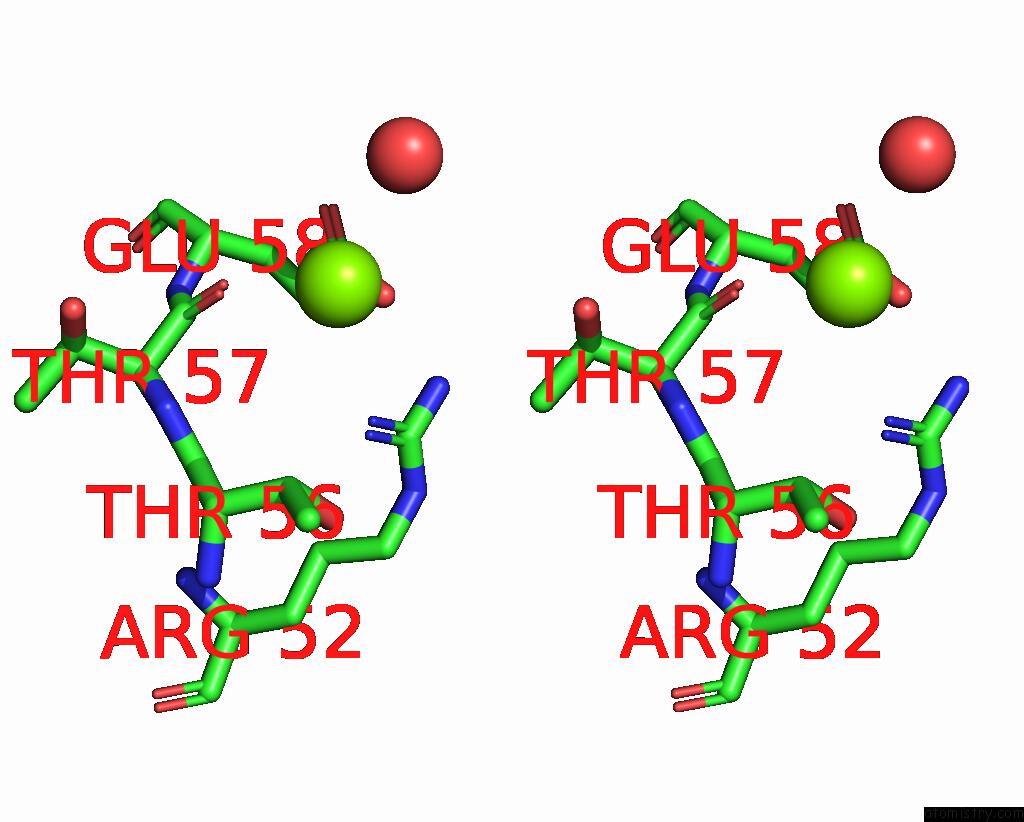
Magnesium binding site 1 out of 2 in 3ijs
Go back to
Magnesium binding site 1 out
of 2 in the Structure of S67-27 in Complex with Tsbp

Mono view
Stereo pair view
Mono view
Stereo pair view
A full contact list of Magnesium with other atoms in the Mg binding
site number 1 of Structure of S67-27 in Complex with Tsbp within 5.0Å range:
|
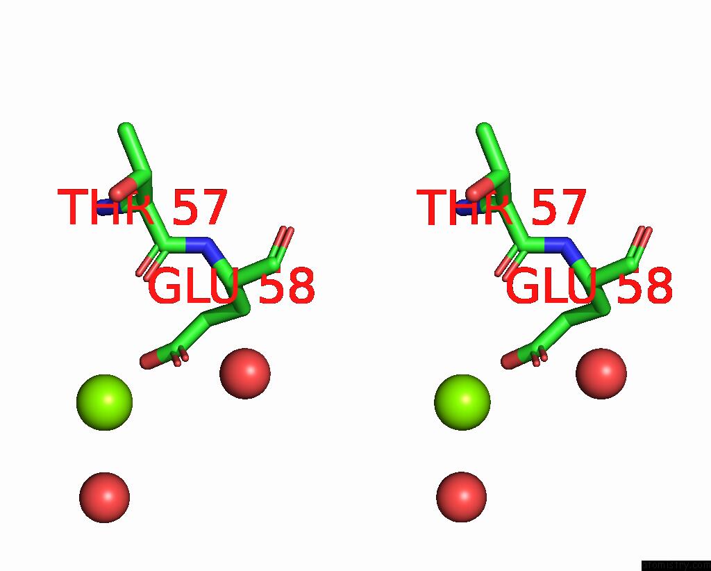
Magnesium binding site 2 out of 2 in 3ijs
Go back to
Magnesium binding site 2 out
of 2 in the Structure of S67-27 in Complex with Tsbp

Mono view
Stereo pair view
Mono view
Stereo pair view
A full contact list of Magnesium with other atoms in the Mg binding
site number 2 of Structure of S67-27 in Complex with Tsbp within 5.0Å range:
|
Reference:
C.L.Brooks,
R.J.Blackler,
G.Sixta,
P.Kosma,
S.Muller-Loennies,
L.Brade,
T.Hirama,
C.R.Mackenzie,
H.Brade,
S.V.Evans.
The Role of Cdr H3 in Antibody Recognition of A Synthetic Analog of A Lipopolysaccharide Antigen. Glycobiology V. 20 138 2010.
ISSN: ISSN 0959-6658
PubMed: 19767317
DOI: 10.1093/GLYCOB/CWP150
Page generated: Sun Aug 10 22:37:36 2025
ISSN: ISSN 0959-6658
PubMed: 19767317
DOI: 10.1093/GLYCOB/CWP150
Last articles
Mg in 6TR4Mg in 6TR3
Mg in 6TMF
Mg in 6TQO
Mg in 6TQN
Mg in 6TQF
Mg in 6TQE
Mg in 6TQB
Mg in 6TQA
Mg in 6TPS