Magnesium »
PDB 3ktv-3l8z »
3l2c »
Magnesium in PDB 3l2c: Crystal Structure of the Dna Binding Domain of FOXO4 Bound to Dna
Protein crystallography data
The structure of Crystal Structure of the Dna Binding Domain of FOXO4 Bound to Dna, PDB code: 3l2c
was solved by
E.Boura,
J.Silhan,
M.Sulc,
J.Brynda,
V.Obsilova,
T.Obsil,
with X-Ray Crystallography technique. A brief refinement statistics is given in the table below:
| Resolution Low / High (Å) | 19.00 / 1.87 |
| Space group | C 2 2 21 |
| Cell size a, b, c (Å), α, β, γ (°) | 40.810, 71.700, 131.860, 90.00, 90.00, 90.00 |
| R / Rfree (%) | 19.3 / 22.8 |
Magnesium Binding Sites:
The binding sites of Magnesium atom in the Crystal Structure of the Dna Binding Domain of FOXO4 Bound to Dna
(pdb code 3l2c). This binding sites where shown within
5.0 Angstroms radius around Magnesium atom.
In total 2 binding sites of Magnesium where determined in the Crystal Structure of the Dna Binding Domain of FOXO4 Bound to Dna, PDB code: 3l2c:
Jump to Magnesium binding site number: 1; 2;
In total 2 binding sites of Magnesium where determined in the Crystal Structure of the Dna Binding Domain of FOXO4 Bound to Dna, PDB code: 3l2c:
Jump to Magnesium binding site number: 1; 2;
Magnesium binding site 1 out of 2 in 3l2c
Go back to
Magnesium binding site 1 out
of 2 in the Crystal Structure of the Dna Binding Domain of FOXO4 Bound to Dna

Mono view
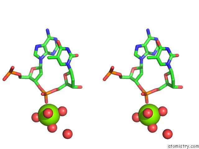
Stereo pair view
Mono view
Stereo pair view
A full contact list of Magnesium with other atoms in the Mg binding
site number 1 of Crystal Structure of the Dna Binding Domain of FOXO4 Bound to Dna within 5.0Å range:
|
Magnesium binding site 2 out of 2 in 3l2c
Go back to
Magnesium binding site 2 out
of 2 in the Crystal Structure of the Dna Binding Domain of FOXO4 Bound to Dna
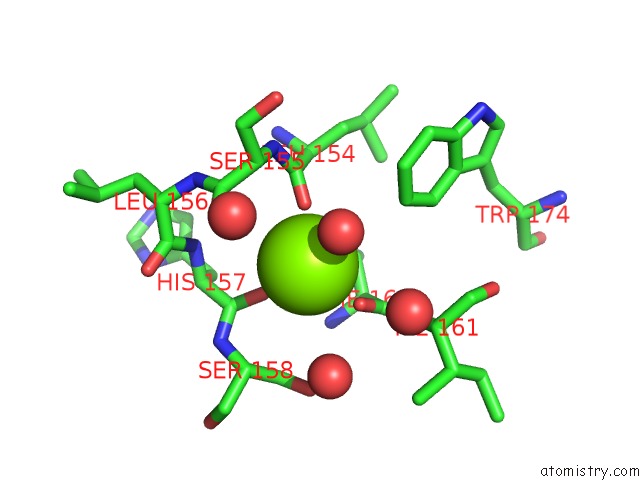
Mono view

Stereo pair view
Mono view
Stereo pair view
A full contact list of Magnesium with other atoms in the Mg binding
site number 2 of Crystal Structure of the Dna Binding Domain of FOXO4 Bound to Dna within 5.0Å range:
|
Reference:
E.Boura,
L.Rezabkova,
J.Brynda,
V.Obsilova,
T.Obsil.
Structure of the Human FOXO4-Dbd-Dna Complex at 1.9 A Resolution Reveals New Details of Foxo Binding to the Dna Acta Crystallogr.,Sect.D V. 66 1351 2010.
ISSN: ISSN 0907-4449
PubMed: 21123876
DOI: 10.1107/S0907444910042228
Page generated: Mon Aug 11 00:01:46 2025
ISSN: ISSN 0907-4449
PubMed: 21123876
DOI: 10.1107/S0907444910042228
Last articles
Mg in 4CYOMg in 4CYJ
Mg in 4CW7
Mg in 4CYN
Mg in 4CYM
Mg in 4CWB
Mg in 4CVN
Mg in 4CT4
Mg in 4CVM
Mg in 4CVO