Magnesium »
PDB 3n8u-3nky »
3nfu »
Magnesium in PDB 3nfu: Crystal Structure of Probable Glucarate Dehydratase From Chromohalobacter Salexigens Dsm 3043 Complexed with Magnesium
Enzymatic activity of Crystal Structure of Probable Glucarate Dehydratase From Chromohalobacter Salexigens Dsm 3043 Complexed with Magnesium
All present enzymatic activity of Crystal Structure of Probable Glucarate Dehydratase From Chromohalobacter Salexigens Dsm 3043 Complexed with Magnesium:
4.2.1.40;
4.2.1.40;
Protein crystallography data
The structure of Crystal Structure of Probable Glucarate Dehydratase From Chromohalobacter Salexigens Dsm 3043 Complexed with Magnesium, PDB code: 3nfu
was solved by
Y.Patskovsky,
R.Toro,
M.Rutter,
J.M.Sauder,
J.A.Gerlt,
S.C.Almo,
S.K.Burley,
New York Structural Genomix Research Consortium(Nysgxrc),
New York Sgx Research Center For Structural Genomics(Nysgxrc),
with X-Ray Crystallography technique. A brief refinement statistics is given in the table below:
| Resolution Low / High (Å) | 20.00 / 1.94 |
| Space group | C 1 2 1 |
| Cell size a, b, c (Å), α, β, γ (°) | 135.009, 105.731, 87.991, 90.00, 128.97, 90.00 |
| R / Rfree (%) | 19.7 / 26.6 |
Magnesium Binding Sites:
The binding sites of Magnesium atom in the Crystal Structure of Probable Glucarate Dehydratase From Chromohalobacter Salexigens Dsm 3043 Complexed with Magnesium
(pdb code 3nfu). This binding sites where shown within
5.0 Angstroms radius around Magnesium atom.
In total 2 binding sites of Magnesium where determined in the Crystal Structure of Probable Glucarate Dehydratase From Chromohalobacter Salexigens Dsm 3043 Complexed with Magnesium, PDB code: 3nfu:
Jump to Magnesium binding site number: 1; 2;
In total 2 binding sites of Magnesium where determined in the Crystal Structure of Probable Glucarate Dehydratase From Chromohalobacter Salexigens Dsm 3043 Complexed with Magnesium, PDB code: 3nfu:
Jump to Magnesium binding site number: 1; 2;
Magnesium binding site 1 out of 2 in 3nfu
Go back to
Magnesium binding site 1 out
of 2 in the Crystal Structure of Probable Glucarate Dehydratase From Chromohalobacter Salexigens Dsm 3043 Complexed with Magnesium
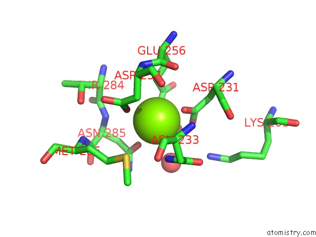
Mono view

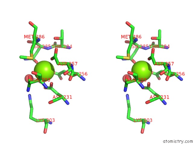
Stereo pair view
Mono view
Stereo pair view
A full contact list of Magnesium with other atoms in the Mg binding
site number 1 of Crystal Structure of Probable Glucarate Dehydratase From Chromohalobacter Salexigens Dsm 3043 Complexed with Magnesium within 5.0Å range:
|
Magnesium binding site 2 out of 2 in 3nfu
Go back to
Magnesium binding site 2 out
of 2 in the Crystal Structure of Probable Glucarate Dehydratase From Chromohalobacter Salexigens Dsm 3043 Complexed with Magnesium

Mono view
Stereo pair view
Mono view
Stereo pair view
A full contact list of Magnesium with other atoms in the Mg binding
site number 2 of Crystal Structure of Probable Glucarate Dehydratase From Chromohalobacter Salexigens Dsm 3043 Complexed with Magnesium within 5.0Å range:
|
Reference:
Y.Patskovsky,
R.Toro,
M.Rutter,
J.M.Sauder,
J.A.Gerlt,
S.K.Burley,
S.C.Almo.
Crystal Structure of Glucarate Dehydratase From Chromohalobacter Salexigens To Be Published.
Page generated: Mon Aug 11 00:53:46 2025
Last articles
Mg in 6IK9Mg in 6IJI
Mg in 6IJH
Mg in 6IID
Mg in 6III
Mg in 6IHW
Mg in 6II6
Mg in 6IHV
Mg in 6IHU
Mg in 6IHS