Magnesium »
PDB 3wqd-3x1l »
3wyi »
Magnesium in PDB 3wyi: Structure of S. Aureus Undecaprenyl Diphosphate Synthase
Protein crystallography data
The structure of Structure of S. Aureus Undecaprenyl Diphosphate Synthase, PDB code: 3wyi
was solved by
J.Gao,
T.P.Ko,
C.H.Huang,
E.Oldfield,
R.T.Guo,
with X-Ray Crystallography technique. A brief refinement statistics is given in the table below:
| Resolution Low / High (Å) | 25.00 / 2.00 |
| Space group | P 31 2 1 |
| Cell size a, b, c (Å), α, β, γ (°) | 62.080, 62.080, 133.486, 90.00, 90.00, 120.00 |
| R / Rfree (%) | 19 / 22.7 |
Magnesium Binding Sites:
The binding sites of Magnesium atom in the Structure of S. Aureus Undecaprenyl Diphosphate Synthase
(pdb code 3wyi). This binding sites where shown within
5.0 Angstroms radius around Magnesium atom.
In total 2 binding sites of Magnesium where determined in the Structure of S. Aureus Undecaprenyl Diphosphate Synthase, PDB code: 3wyi:
Jump to Magnesium binding site number: 1; 2;
In total 2 binding sites of Magnesium where determined in the Structure of S. Aureus Undecaprenyl Diphosphate Synthase, PDB code: 3wyi:
Jump to Magnesium binding site number: 1; 2;
Magnesium binding site 1 out of 2 in 3wyi
Go back to
Magnesium binding site 1 out
of 2 in the Structure of S. Aureus Undecaprenyl Diphosphate Synthase
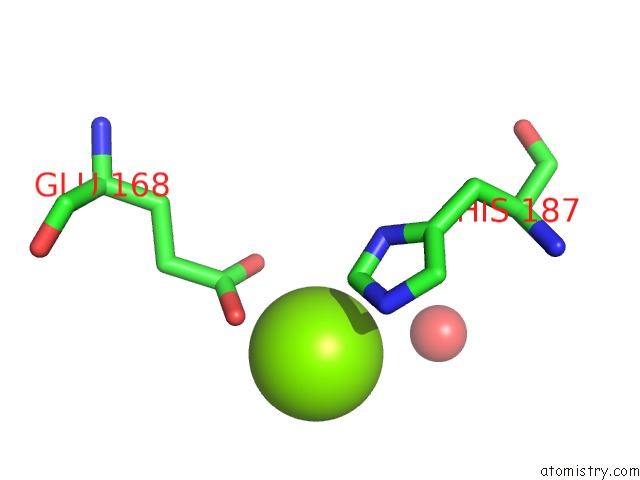
Mono view
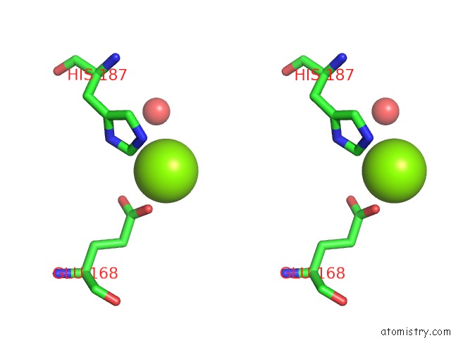
Stereo pair view
Mono view
Stereo pair view
A full contact list of Magnesium with other atoms in the Mg binding
site number 1 of Structure of S. Aureus Undecaprenyl Diphosphate Synthase within 5.0Å range:
|
Magnesium binding site 2 out of 2 in 3wyi
Go back to
Magnesium binding site 2 out
of 2 in the Structure of S. Aureus Undecaprenyl Diphosphate Synthase

Mono view

Stereo pair view
Mono view
Stereo pair view
A full contact list of Magnesium with other atoms in the Mg binding
site number 2 of Structure of S. Aureus Undecaprenyl Diphosphate Synthase within 5.0Å range:
|
Reference:
W.Zhu,
Y.Wang,
K.Li,
J.Gao,
C.H.Huang,
C.C.Chen,
T.P.Ko,
Y.Zhang,
R.T.Guo,
E.Oldfield.
Antibacterial Drug Leads: Dna and Enzyme Multitargeting. J.Med.Chem. 2015.
ISSN: ISSN 0022-2623
PubMed: 25574764
DOI: 10.1021/JM501449U
Page generated: Mon Aug 11 05:04:52 2025
ISSN: ISSN 0022-2623
PubMed: 25574764
DOI: 10.1021/JM501449U
Last articles
Mg in 6KOUMg in 6KPE
Mg in 6KOQ
Mg in 6KNZ
Mg in 6KOP
Mg in 6KON
Mg in 6KOO
Mg in 6KO6
Mg in 6KO1
Mg in 6KMA