Magnesium »
PDB 4brs-4c2z »
4c06 »
Magnesium in PDB 4c06: Crystal Structure of M. Musculus Protein Arginine Methyltransferase PRMT6 with MGCL2
Enzymatic activity of Crystal Structure of M. Musculus Protein Arginine Methyltransferase PRMT6 with MGCL2
All present enzymatic activity of Crystal Structure of M. Musculus Protein Arginine Methyltransferase PRMT6 with MGCL2:
2.1.1.125;
2.1.1.125;
Protein crystallography data
The structure of Crystal Structure of M. Musculus Protein Arginine Methyltransferase PRMT6 with MGCL2, PDB code: 4c06
was solved by
L.Bonnefond,
V.Cura,
N.Troffer-Charlier,
J.Mailliot,
J.M.Wurtz,
J.Cavarelli,
with X-Ray Crystallography technique. A brief refinement statistics is given in the table below:
| Resolution Low / High (Å) | 40.923 / 1.60 |
| Space group | I 41 |
| Cell size a, b, c (Å), α, β, γ (°) | 79.758, 79.758, 118.945, 90.00, 90.00, 90.00 |
| R / Rfree (%) | 15.54 / 19.39 |
Magnesium Binding Sites:
The binding sites of Magnesium atom in the Crystal Structure of M. Musculus Protein Arginine Methyltransferase PRMT6 with MGCL2
(pdb code 4c06). This binding sites where shown within
5.0 Angstroms radius around Magnesium atom.
In total only one binding site of Magnesium was determined in the Crystal Structure of M. Musculus Protein Arginine Methyltransferase PRMT6 with MGCL2, PDB code: 4c06:
In total only one binding site of Magnesium was determined in the Crystal Structure of M. Musculus Protein Arginine Methyltransferase PRMT6 with MGCL2, PDB code: 4c06:
Magnesium binding site 1 out of 1 in 4c06
Go back to
Magnesium binding site 1 out
of 1 in the Crystal Structure of M. Musculus Protein Arginine Methyltransferase PRMT6 with MGCL2
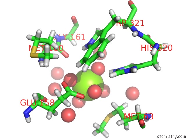
Mono view
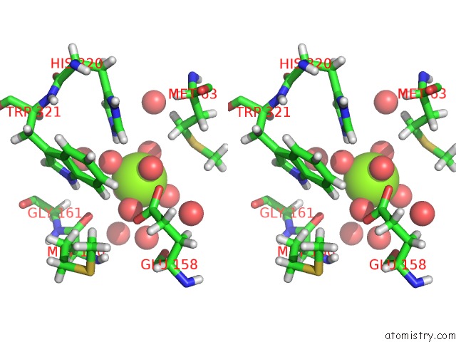
Stereo pair view
Mono view
Stereo pair view
A full contact list of Magnesium with other atoms in the Mg binding
site number 1 of Crystal Structure of M. Musculus Protein Arginine Methyltransferase PRMT6 with MGCL2 within 5.0Å range:
|
Reference:
L.Bonnefond,
V.Cura,
N.Troffer-Charlier,
J.Mailliot,
J.M.Wurtz,
J.Cavarelli.
Crystal Structure of M. Musculus Protein Arginine Methyltransferase PRMT6 To Be Published.
Page generated: Mon Aug 11 07:08:51 2025
Last articles
Mg in 4ZK5Mg in 4ZJJ
Mg in 4ZJI
Mg in 4ZK4
Mg in 4ZIR
Mg in 4ZIY
Mg in 4ZIB
Mg in 4ZI7
Mg in 4ZIL
Mg in 4ZI5