Magnesium »
PDB 4brs-4c2z »
4c0l »
Magnesium in PDB 4c0l: Crystal Structure of Drosophila Miro Ef Hand and Cgtpase Domains Bound to One Magnesium Ion and Mg:Gdp (Mggdp-Miros)
Protein crystallography data
The structure of Crystal Structure of Drosophila Miro Ef Hand and Cgtpase Domains Bound to One Magnesium Ion and Mg:Gdp (Mggdp-Miros), PDB code: 4c0l
was solved by
J.L.Klosowiak,
P.J.Focia,
Z.Wawrzak,
S.Chakravarthy,
E.C.Landahl,
D.M.Freymann,
S.E.Rice,
with X-Ray Crystallography technique. A brief refinement statistics is given in the table below:
| Resolution Low / High (Å) | 25.825 / 3.00 |
| Space group | P 32 2 1 |
| Cell size a, b, c (Å), α, β, γ (°) | 81.510, 81.510, 154.950, 90.00, 90.00, 120.00 |
| R / Rfree (%) | 21.45 / 25.85 |
Other elements in 4c0l:
The structure of Crystal Structure of Drosophila Miro Ef Hand and Cgtpase Domains Bound to One Magnesium Ion and Mg:Gdp (Mggdp-Miros) also contains other interesting chemical elements:
| Sodium | (Na) | 1 atom |
Magnesium Binding Sites:
The binding sites of Magnesium atom in the Crystal Structure of Drosophila Miro Ef Hand and Cgtpase Domains Bound to One Magnesium Ion and Mg:Gdp (Mggdp-Miros)
(pdb code 4c0l). This binding sites where shown within
5.0 Angstroms radius around Magnesium atom.
In total 2 binding sites of Magnesium where determined in the Crystal Structure of Drosophila Miro Ef Hand and Cgtpase Domains Bound to One Magnesium Ion and Mg:Gdp (Mggdp-Miros), PDB code: 4c0l:
Jump to Magnesium binding site number: 1; 2;
In total 2 binding sites of Magnesium where determined in the Crystal Structure of Drosophila Miro Ef Hand and Cgtpase Domains Bound to One Magnesium Ion and Mg:Gdp (Mggdp-Miros), PDB code: 4c0l:
Jump to Magnesium binding site number: 1; 2;
Magnesium binding site 1 out of 2 in 4c0l
Go back to
Magnesium binding site 1 out
of 2 in the Crystal Structure of Drosophila Miro Ef Hand and Cgtpase Domains Bound to One Magnesium Ion and Mg:Gdp (Mggdp-Miros)
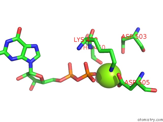
Mono view
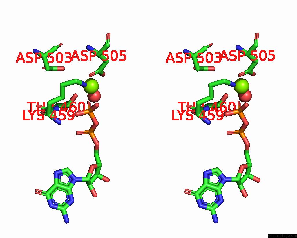
Stereo pair view
Mono view
Stereo pair view
A full contact list of Magnesium with other atoms in the Mg binding
site number 1 of Crystal Structure of Drosophila Miro Ef Hand and Cgtpase Domains Bound to One Magnesium Ion and Mg:Gdp (Mggdp-Miros) within 5.0Å range:
|
Magnesium binding site 2 out of 2 in 4c0l
Go back to
Magnesium binding site 2 out
of 2 in the Crystal Structure of Drosophila Miro Ef Hand and Cgtpase Domains Bound to One Magnesium Ion and Mg:Gdp (Mggdp-Miros)
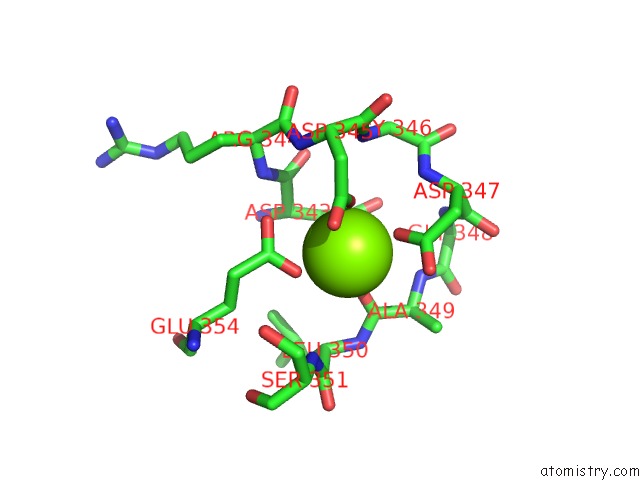
Mono view
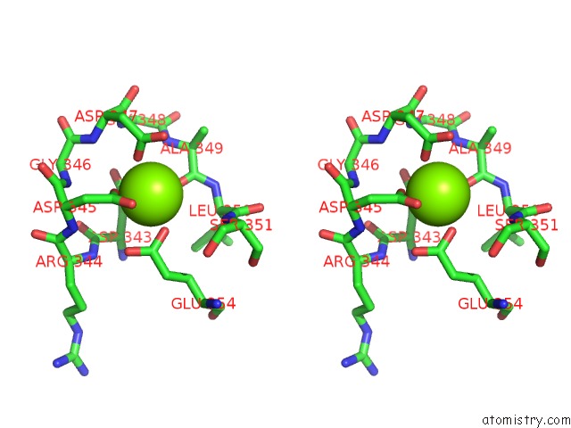
Stereo pair view
Mono view
Stereo pair view
A full contact list of Magnesium with other atoms in the Mg binding
site number 2 of Crystal Structure of Drosophila Miro Ef Hand and Cgtpase Domains Bound to One Magnesium Ion and Mg:Gdp (Mggdp-Miros) within 5.0Å range:
|
Reference:
J.L.Klosowiak,
P.J.Focia,
S.Chakravarthy,
E.C.Landahl,
D.M.Freymann,
S.E.Rice.
Structural Coupling of the Ef Hand and C-Terminal Gtpase Domains in the Mitochondrial Protein Miro. Embo Rep. V. 14 968 2013.
ISSN: ISSN 1469-221X
PubMed: 24071720
DOI: 10.1038/EMBOR.2013.151
Page generated: Mon Aug 11 07:08:53 2025
ISSN: ISSN 1469-221X
PubMed: 24071720
DOI: 10.1038/EMBOR.2013.151
Last articles
Mg in 4Z6CMg in 4Z6D
Mg in 4Z5Q
Mg in 4Z54
Mg in 4Z5Z
Mg in 4Z53
Mg in 4Z4Q
Mg in 4Z4H
Mg in 4Z4I
Mg in 4Z51